3. FOOD ChEMISTRy & bIOTEChNOLOGy 3.1. Lectures
3. FOOD ChEMISTRy & bIOTEChNOLOGy 3.1. Lectures
3. FOOD ChEMISTRy & bIOTEChNOLOGy 3.1. Lectures

Create successful ePaper yourself
Turn your PDF publications into a flip-book with our unique Google optimized e-Paper software.
Chem. Listy, 102, s265–s1311 (2008) Food Chemistry & Biotechnology<br />
identically as described in our previous papers, involving the<br />
DPPH, TBARS and FRP assays. Total phenolic compounds<br />
content of extracts was evaluated, as well. 9, 11<br />
M u l t i v a r i a t e S t a t i s t i c a l A n a l y s i s<br />
Canonical discriminant analysis of all results obtained<br />
from UV-VIS experiments was performed using the Unistat ®<br />
software in order to distinguish the native (non-irradiated)<br />
spice samples from that exposed to γ-radiation.<br />
Results<br />
Microbiological analysis performed immediately after<br />
the irradiation process proved, that as a result of γ-irradiation,<br />
the total count of microorganisms in caraway sample irradiated<br />
at dose of 5kGy decreased considerably from 2.8 × 10 4<br />
colony forming unit (CFU) detected in reference sample, to<br />
less than 10 CFU g –1 . The same effect of γ-irradiation on laurel<br />
leaves was achieved using the dose of 10 kGy, still fulfilling<br />
the requirements of international standards on irradiation<br />
3,4 .<br />
Table I<br />
Microbiological analysis of ground caraway seeds (C) and<br />
laurel bay leaves (L) samples, γ-irradiated at doses of <br />
kGy using 60Co-source performed one day after the γ-irradiation<br />
Radiation Total count of<br />
dose microorganisms<br />
Coliform<br />
bacteria<br />
Moulds<br />
[CFU g –1 [kGy] [CFU g<br />
]<br />
–1 ] [CFU g –1 ]<br />
C L C L C L<br />
0 2.8 × 10 4 1.7 × 10 5 1.0 × 10 4 8.6 × 10 3 9.8 × 10 3 5.7 × 10 3<br />
5 < 10 5.7 × 10 3 < 10 5.0 × 10 1 2.5 × 10 2 < 10<br />
10 < 10 < 10 < 10 < 10 < 10 < 10<br />
20 < 10 < 10 < 10 < 10 < 10 < 10<br />
30 < 10 < 10 < 10 < 10 < 10 < 10<br />
As follows from data presented in Table I, the presence of<br />
oliform bacteria as well as of yields and moulds was effectively<br />
suppressed by the irradiation. Analysis performed 6 months of<br />
post-irradiation storage confirmed, that microbial status of both<br />
spices remained practically unchanged.<br />
EPR spectrum of both reference (non-irradiated) samples<br />
represents broad singlet line with unresolved hyperfine splitting,<br />
attributable mostly to Mn 2+ ions, upon which the additional sharp<br />
EPR line (g eff = 2.0022, ∆B pp ~ 1 mT) is superimposed, previously<br />
assigned to stable semiquinone radicals produced by the<br />
oxidation of polyphenolic compounds present in plants. In addition,<br />
the presence of low-intensive EPR singlet line was noticed<br />
in caraway reference sample, attributable to radicals generated<br />
during the grinding process (Table II). 5,9–11<br />
EPR spectra of γ-radiation treated spices showed the formation<br />
of additional paramagnetic structures. As follows from<br />
detail simulation analysis of obtained spectra (Table II), different,<br />
mostly cellulosic and carbohydrate radical structures were<br />
identified.<br />
s561<br />
Table II<br />
Identification of radical structures found in reference and<br />
γ-irradiated samples of ground caraway and laurel leaves<br />
EPR signal g-value Hyperfine ΔB pp [mT]<br />
origin splittings [mT]<br />
Reference samples<br />
Semiquinones g ┴ = 2.0042 – 0.52<br />
g ║ = 2.0030<br />
Carbohydrate I g ┴ = 2.0041 – 0.06<br />
g ║ = 2.0028<br />
γ- irradiated samples<br />
Carbohydrate II g ┴ = 2.0041 A ┴ = 0.7 0.45<br />
g ║ = 2.0033 A ║ = 0.6 (2H)<br />
Carbohydrate III g ┴ = 2.0032 A ┴ = 0.85 0.67<br />
g ║ = 2.0025 A ║ = 0.7 (2H)<br />
Carbohydrate IV g ┴ = 2.0030 A ┴ = 0.45 0.59<br />
g ║ = 2.0038 A ║ = 0.40 (1H)<br />
Cellulosic g ┴ = 2.0029 A ┴ = <strong>3.</strong>00 1.20<br />
g ║ = 2.0014 A ║ = 1.70 (2H)<br />
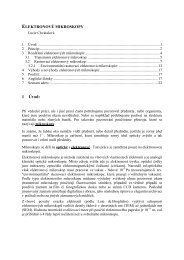
Integral EPR intensity<br />
8<br />
7<br />
6<br />
5<br />
4<br />
3<br />
2<br />
1<br />
March<br />
April<br />
May<br />
June<br />
0 5 10 15 20 25 30<br />
Radiation dose, kGy<br />
Fig. 1. Dependence of integral EPR intensity of ground caraway<br />
seed on γ-radiation dose measured immediately after<br />
the irradiation (March) and during three month of post irradiation<br />
storage (April-june). EPR spectra were recorded using<br />
0.633 mw microwave power at 298 K<br />
These radicals originate either from cleavage processes<br />
of cellulose matter (laurel leaves) and/or of other polysaccharides<br />
forming the skeleton of plant structures and their cells,<br />
as the cellulosic radicals were not detected in γ-irradiated caraway<br />
samples.<br />
In accord with our previously published papers, the<br />
dose-dependent formation of radical structures’ in γ-irradiated<br />
samples of both spices under study was found (Fig. 1.).