Noncontact Atomic Force Microscopy - Yale School of Engineering ...
Noncontact Atomic Force Microscopy - Yale School of Engineering ...
Noncontact Atomic Force Microscopy - Yale School of Engineering ...

Create successful ePaper yourself
Turn your PDF publications into a flip-book with our unique Google optimized e-Paper software.
P.II-18<br />
Development <strong>of</strong> a NC – AFM for Ambient and Liquid Environments<br />
Haider I. Rasool 1 , Shivani Sharma 1 , James K. Gimzewski 1,2,3<br />
1 Department <strong>of</strong> Chemistry and Biochemistry, University <strong>of</strong> California – Los Angeles, USA<br />
2 California NanoSystems Institute, 570 Westwood Plaza, Los Angeles, USA.<br />
3 International Center for Materials Nanoarchitectonics (MANA), Tsukuba, Japan<br />
High resolution Non – Contact <strong>Atomic</strong> <strong>Force</strong> <strong>Microscopy</strong> imaging has been<br />
achieved by various groups in both ambient and liquid environments [1] – [3]. The recent<br />
success <strong>of</strong> NC – AFM in these “low – Q” environments has been attributable to the<br />
development <strong>of</strong> low noise detection schemes for measuring small amplitude deflections.<br />
We describe current research on the development <strong>of</strong> a robust home – built scanning probe<br />
microscope with an incorporated low noise all fiber interferometer deflection sensor for<br />
imaging in both ambient and liquid environments. The current design <strong>of</strong> the scanning<br />
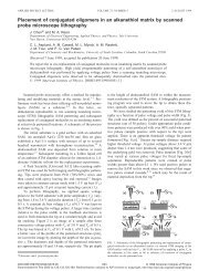
probe microscope in its AFM configuration is depicted in Figure 1 (a).<br />
The developing instrument has been used to image various biological species in<br />
both ambient and liquid environments in different imaging modes. In particular, our<br />
group has been successful in imaging isolated human saliva exosomes fixed to a mica<br />
substrate in both air and buffer solution. Both amplitude and phase modulation imaging<br />
have been accomplished on these samples under ambient conditions. Using constant<br />
excitation phase modulated imaging, a simultaneous mapping <strong>of</strong> topography and energy<br />
dissipation has been possible at a constant average tip sample force. Figure 1(b) shows a<br />
typical image <strong>of</strong> the topography <strong>of</strong> an isolated exosome.<br />
a) b)<br />
Figure 1: The above images show (a) the design <strong>of</strong> the home built microscope and (b) a PM –<br />
AFM image <strong>of</strong> a human saliva exosome.<br />
[1] T. Fukuma, M. Kimura, K. Kobayashi, K. Matsushige, and H. Yamada. Rev. Sci. Instrum. 76, 053704<br />
(2005).<br />
[2] B.W. Hoogenboom, H.J. Hug, Y. Pellmont, S. Martin, P.L.T.M. Frederix, D. Fotiadis,and A. Engel.<br />
Appl. Phys. Lett. 88, 193109 (2006).<br />
[3] T. Fukuma, and S. Jarvis. Rev. Sci. Instrum. 77, 043701 (2006).<br />
146