2012 COURSE DATES: AUGUST 4 – 17, 2012 - Sirenian International
2012 COURSE DATES: AUGUST 4 – 17, 2012 - Sirenian International
2012 COURSE DATES: AUGUST 4 – 17, 2012 - Sirenian International

You also want an ePaper? Increase the reach of your titles
YUMPU automatically turns print PDFs into web optimized ePapers that Google loves.
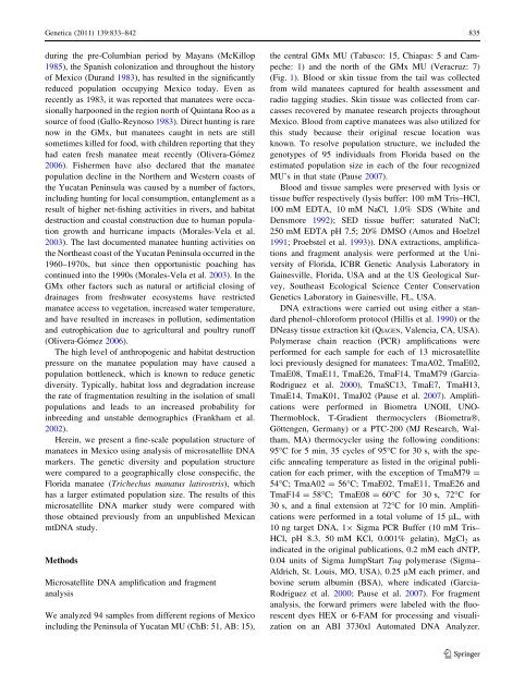
Genetica (2011) 139:833<strong>–</strong>842 835<br />
during the pre-Columbian period by Mayans (McKillop<br />
1985), the Spanish colonization and throughout the history<br />
of Mexico (Durand 1983), has resulted in the significantly<br />
reduced population occupying Mexico today. Even as<br />
recently as 1983, it was reported that manatees were occasionally<br />
harpooned in the region north of Quintana Roo as a<br />
source of food (Gallo-Reynoso 1983). Direct hunting is rare<br />
now in the GMx, but manatees caught in nets are still<br />
sometimes killed for food, with children reporting that they<br />
had eaten fresh manatee meat recently (Olivera-Gómez<br />
2006). Fishermen have also declared that the manatee<br />
population decline in the Northern and Western coasts of<br />
the Yucatan Peninsula was caused by a number of factors,<br />
including hunting for local consumption, entanglement as a<br />
result of higher net-fishing activities in rivers, and habitat<br />
destruction and coastal construction due to human population<br />
growth and hurricane impacts (Morales-Vela et al.<br />
2003). The last documented manatee hunting activities on<br />
the Northeast coast of the Yucatan Peninsula occurred in the<br />
1960<strong>–</strong>1970s, but since then opportunistic poaching has<br />
continued into the 1990s (Morales-Vela et al. 2003). In the<br />
GMx other factors such as natural or artificial closing of<br />
drainages from freshwater ecosystems have restricted<br />
manatee access to vegetation, increased water temperature,<br />
and have resulted in increases in pollution, sedimentation<br />
and eutrophication due to agricultural and poultry runoff<br />
(Olivera-Gómez 2006).<br />
The high level of anthropogenic and habitat destruction<br />
pressure on the manatee population may have caused a<br />
population bottleneck, which is known to reduce genetic<br />
diversity. Typically, habitat loss and degradation increase<br />
the rate of fragmentation resulting in the isolation of small<br />
populations and leads to an increased probability for<br />
inbreeding and unstable demographics (Frankham et al.<br />
2002).<br />
Herein, we present a fine-scale population structure of<br />
manatees in Mexico using analysis of microsatellite DNA<br />
markers. The genetic diversity and population structure<br />
were compared to a geographically close conspecific, the<br />
Florida manatee (Trichechus manatus latirostris), which<br />
has a larger estimated population size. The results of this<br />
microsatellite DNA marker study were compared with<br />
those obtained previously from an unpublished Mexican<br />
mtDNA study.<br />
Methods<br />
Microsatellite DNA amplification and fragment<br />
analysis<br />
We analyzed 94 samples from different regions of Mexico<br />
including the Peninsula of Yucatan MU (ChB: 51, AB: 15),<br />
the central GMx MU (Tabasco: 15, Chiapas: 5 and Campeche:<br />
1) and the north of the GMx MU (Veracruz: 7)<br />
(Fig. 1). Blood or skin tissue from the tail was collected<br />
from wild manatees captured for health assessment and<br />
radio tagging studies. Skin tissue was collected from carcasses<br />
recovered by manatee research projects throughout<br />
Mexico. Blood from captive manatees was also utilized for<br />
this study because their original rescue location was<br />
known. To resolve population structure, we included the<br />
genotypes of 95 individuals from Florida based on the<br />
estimated population size in each of the four recognized<br />
MU’s in that state (Pause 2007).<br />
Blood and tissue samples were preserved with lysis or<br />
tissue buffer respectively (lysis buffer: 100 mM Tris<strong>–</strong>HCl,<br />
100 mM EDTA, 10 mM NaCl, 1.0% SDS (White and<br />
Densmore 1992); SED tissue buffer: saturated NaCl;<br />
250 mM EDTA pH 7.5; 20% DMSO (Amos and Hoelzel<br />
1991; Proebstel et al. 1993)). DNA extractions, amplifications<br />
and fragment analysis were performed at the University<br />
of Florida, ICBR Genetic Analysis Laboratory in<br />
Gainesville, Florida, USA and at the US Geological Survey,<br />
Southeast Ecological Science Center Conservation<br />
Genetics Laboratory in Gainesville, FL, USA.<br />
DNA extractions were carried out using either a standard<br />
phenol<strong>–</strong>chloroform protocol (Hillis et al. 1990) or the<br />
DNeasy tissue extraction kit (QIAGEN, Valencia, CA, USA).<br />
Polymerase chain reaction (PCR) amplifications were<br />
performed for each sample for each of 13 microsatellite<br />
loci previously designed for manatees: TmaA02, TmaE02,<br />
TmaE08, TmaE11, TmaE26, TmaF14, TmaM79 (Garcia-<br />
Rodriguez et al. 2000), TmaSC13, TmaE7, TmaH13,<br />
TmaE14, TmaK01, TmaJ02 (Pause et al. 2007). Amplifications<br />
were performed in Biometra UNOII, UNO-<br />
Thermoblock, T-Gradient thermocyclers (BiometraÒ,<br />
Göttengen, Germany) or a PTC-200 (MJ Research, Waltham,<br />
MA) thermocycler using the following conditions:<br />
95°C for 5 min, 35 cycles of 95°C for 30 s, with the specific<br />
annealing temperature as listed in the original publication<br />
for each primer, with the exception of TmaM79 =<br />
54°C; TmaA02 = 56°C; TmaE02, TmaE11, TmaE26 and<br />
TmaF14 = 58°C; TmaE08 = 60°C for 30 s, 72°C for<br />
30 s, and a final extension at 72°C for 10 min. Amplifications<br />
were performed in a total volume of 15 lL, with<br />
10 ng target DNA, 19 Sigma PCR Buffer (10 mM Tris<strong>–</strong><br />
HCl, pH 8.3, 50 mM KCl, 0.001% gelatin), MgCl2 as<br />
indicated in the original publications, 0.2 mM each dNTP,<br />
0.04 units of Sigma JumpStart Taq polymerase (Sigma<strong>–</strong><br />
Aldrich, St. Louis, MO, USA), 0.25 lM each primer, and<br />
bovine serum albumin (BSA), where indicated (Garcia-<br />
Rodriguez et al. 2000; Pause et al. 2007). For fragment<br />
analysis, the forward primers were labeled with the fluorescent<br />
dyes HEX or 6-FAM for processing and visualization<br />
on an ABI 3730xl Automated DNA Analyzer.<br />
123