Chapter 1, The Reptilian Spectacle - UWSpace - University of ...
Chapter 1, The Reptilian Spectacle - UWSpace - University of ...
Chapter 1, The Reptilian Spectacle - UWSpace - University of ...

You also want an ePaper? Increase the reach of your titles
YUMPU automatically turns print PDFs into web optimized ePapers that Google loves.
quote above, the relationship between that shed scale and the eye remained open to debate and<br />
speculation on whether the scale was affixed to the cornea, to an eyelid, or if it simply floated over the<br />
cornea (reviewed in Cloquet 1821).<br />
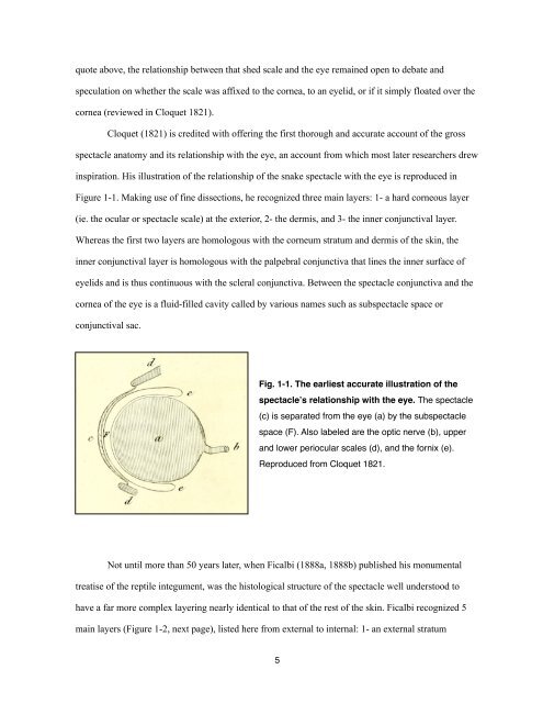
Cloquet (1821) is credited with <strong>of</strong>fering the first thorough and accurate account <strong>of</strong> the gross<br />
spectacle anatomy and its relationship with the eye, an account from which most later researchers drew<br />
inspiration. His illustration <strong>of</strong> the relationship <strong>of</strong> the snake spectacle with the eye is reproduced in<br />
Figure 1-1. Making use <strong>of</strong> fine dissections, he recognized three main layers: 1- a hard corneous layer<br />
(ie. the ocular or spectacle scale) at the exterior, 2- the dermis, and 3- the inner conjunctival layer.<br />
Whereas the first two layers are homologous with the corneum stratum and dermis <strong>of</strong> the skin, the<br />
inner conjunctival layer is homologous with the palpebral conjunctiva that lines the inner surface <strong>of</strong><br />
eyelids and is thus continuous with the scleral conjunctiva. Between the spectacle conjunctiva and the<br />
cornea <strong>of</strong> the eye is a fluid-filled cavity called by various names such as subspectacle space or<br />
conjunctival sac.<br />
Fig. 1-1. <strong>The</strong> earliest accurate illustration <strong>of</strong> the<br />
spectacleʼs relationship with the eye. <strong>The</strong> spectacle<br />
(c) is separated from the eye (a) by the subspectacle<br />
space (F). Also labeled are the optic nerve (b), upper<br />
and lower periocular scales (d), and the fornix (e).<br />
Reproduced from Cloquet 1821.<br />
Not until more than 50 years later, when Ficalbi (1888a, 1888b) published his monumental<br />
treatise <strong>of</strong> the reptile integument, was the histological structure <strong>of</strong> the spectacle well understood to<br />
have a far more complex layering nearly identical to that <strong>of</strong> the rest <strong>of</strong> the skin. Ficalbi recognized 5<br />
main layers (Figure 1-2, next page), listed here from external to internal: 1- an external stratum<br />
5