- Page 1 and 2: Investigations on the Reptilian Spe
- Page 3 and 4: Abstract The eyes of snakes and mos
- Page 5 and 6: Heritage Library, Smithsonian Libra
- Page 7 and 8: 2.2.1 Animals 36 2.2.2 Experimental
- Page 9 and 10: List of Figures 1-1. The earliest a
- Page 11 and 12: List of Tables 2-1. Durations of sp
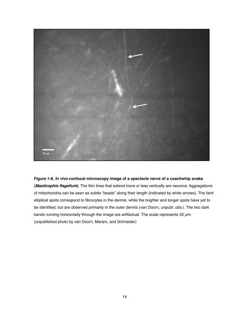
- Page 13 and 14: UV-A Ultraviolet A (315-400 nm) UV-
- Page 15 and 16: “Il est de connaissance presque v
- Page 17 and 18: 1.1 Anatomy of the spectacle The si
- Page 19 and 20: corneum, 2- an inner stratum corneu
- Page 21 and 22: The two layers he described in the
- Page 23 and 24: Figure 1-5. Illustration of the spe
- Page 25: injected spectacles clearly shows t
- Page 29 and 30: 1.1.3 The Spectacles of Geckos Most
- Page 31 and 32: 1.1.4 The Spectacles of Amphisbaeni
- Page 33 and 34: Gymnophthalmus (spectacled tegus) a
- Page 35 and 36: appreciates on observing the sadly
- Page 37 and 38: hydration state (van Doorn, unpubl.
- Page 39 and 40: 1.5 Adaptive Significance of the Sp
- Page 41 and 42: diurnally active in hot and dry hab
- Page 43 and 44: of his argument hinges on the prese
- Page 45 and 46: • In Chapter 3, findings on the v
- Page 47 and 48: 2.1 Introduction This introduction
- Page 49 and 50: 2.2 Methods & Materials The experim
- Page 51 and 52: To allow for extended high-magnific
- Page 53 and 54: At 30 minutes into the experiment,
- Page 55 and 56: packages. All statistical analyses
- Page 57 and 58: 2.3 Results 2.3.1 Spectacle blood f
- Page 59 and 60: Figure 2-7. Graph of two representa
- Page 61 and 62: 2.4 Discussion The purpose of this
- Page 63 and 64: vascular changes were occurring onl
- Page 65 and 66: Chapter 3, Spectral Transmission of
- Page 67 and 68: like the cornea, may exhibit simila
- Page 69 and 70: The scales of both the right and le
- Page 71 and 72: Figure 3-1. Spectral transmittance
- Page 73 and 74: % Transmittance % Transmission % Tr
- Page 75 and 76: % % Transmittance Transmission % %
- Page 77 and 78:
λ 50% (nm) 420 400 380 360 340 320
- Page 79 and 80:
Thickness (!m) Figure 3-7. Plot of
- Page 81 and 82:
Family Spearmanʼs rho p Colubridae
- Page 83 and 84:
virtue of both being characteristic
- Page 85 and 86:
nocturnal (Brattstrom 1952; Klauber
- Page 87 and 88:
Another potential factor that may g
- Page 89 and 90:
Colubridae Colubrinae Elaphe taeniu
- Page 91 and 92:
Pythonidae Python sebae Rock Python
- Page 93 and 94:
4.1 Introduction The reptilian spec
- Page 95 and 96:
thus be optimized to meet not only
- Page 97 and 98:
4.2 Methods & Materials 4.2.1 Sampl
- Page 99 and 100:
for 15 minutes to remove all residu
- Page 101 and 102:
4.3 Results 4.3.1 Keratins of Coach
- Page 103 and 104:
24 kDa > 17 kDa > 12 kDa > S1 P1 V1
- Page 105 and 106:
76 kDa > 52 kDa > 24 kDa > 17 kDa >
- Page 107 and 108:
31 kDa > 24 kDa > 17 kDa > 12 kDa >
- Page 109 and 110:
4.3.4 2D Electrophoretic Comparison
- Page 111 and 112:
kDa 76 - 52 - 38 - 31 - 24 - 17 - 1
- Page 113 and 114:
(2007a) have theorized that basic
- Page 115 and 116:
spectacle, this layer may be partic
- Page 117 and 118:
Chapter 5, Summary and Concluding R
- Page 119 and 120:
evaluations of the elastic modulus
- Page 121 and 122:
References Addison WHF, How HW. 192
- Page 123 and 124:
Berkley MA, Wakins DW. 1973. Gratin
- Page 125 and 126:
Chou BR, Hawryshyn CW. 1987. Spectr
- Page 127 and 128:
Fox RH, Edholm OG. 1963. Nervous co
- Page 129 and 130:
Hinkley JA, Savitsky AH, River G, G
- Page 131 and 132:
Lee P, Wang CC, Adamis AP. 1998. Oc
- Page 133 and 134:
Mautz WJ. 1982. Patterns of evapora
- Page 135 and 136:
Rice GE, Bradshaw SD. 1980. Changes
- Page 137 and 138:
Sillman AJ, Govardovskiĭ WI, Röhl
- Page 139 and 140:
Valentin G. 1879a. Ein Beitrag zur
- Page 141 and 142:
Appendix A One cycle of spectacle b