Chapter 1, The Reptilian Spectacle - UWSpace - University of ...
Chapter 1, The Reptilian Spectacle - UWSpace - University of ...
Chapter 1, The Reptilian Spectacle - UWSpace - University of ...

You also want an ePaper? Increase the reach of your titles
YUMPU automatically turns print PDFs into web optimized ePapers that Google loves.
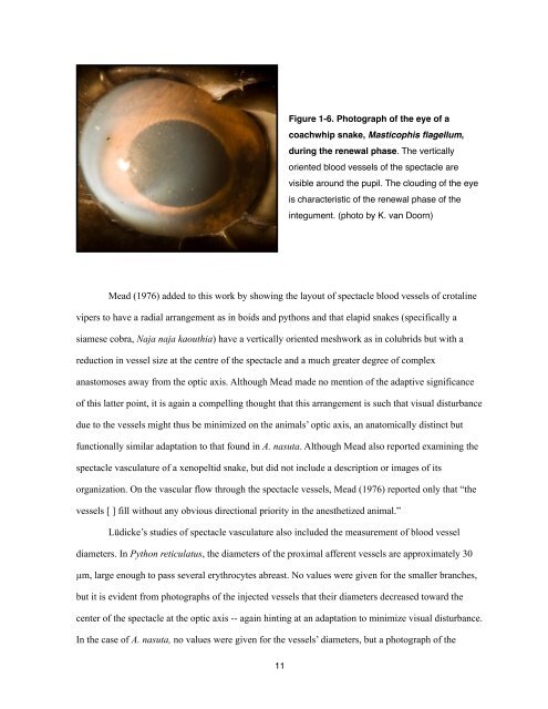
Figure 1-6. Photograph <strong>of</strong> the eye <strong>of</strong> a<br />
coachwhip snake, Masticophis flagellum,<br />
during the renewal phase. <strong>The</strong> vertically<br />
oriented blood vessels <strong>of</strong> the spectacle are<br />
visible around the pupil. <strong>The</strong> clouding <strong>of</strong> the eye<br />
is characteristic <strong>of</strong> the renewal phase <strong>of</strong> the<br />
integument. (photo by K. van Doorn)<br />
Mead (1976) added to this work by showing the layout <strong>of</strong> spectacle blood vessels <strong>of</strong> crotaline<br />
vipers to have a radial arrangement as in boids and pythons and that elapid snakes (specifically a<br />
siamese cobra, Naja naja kaouthia) have a vertically oriented meshwork as in colubrids but with a<br />
reduction in vessel size at the centre <strong>of</strong> the spectacle and a much greater degree <strong>of</strong> complex<br />
anastomoses away from the optic axis. Although Mead made no mention <strong>of</strong> the adaptive significance<br />
<strong>of</strong> this latter point, it is again a compelling thought that this arrangement is such that visual disturbance<br />
due to the vessels might thus be minimized on the animals’ optic axis, an anatomically distinct but<br />
functionally similar adaptation to that found in A. nasuta. Although Mead also reported examining the<br />
spectacle vasculature <strong>of</strong> a xenopeltid snake, but did not include a description or images <strong>of</strong> its<br />
organization. On the vascular flow through the spectacle vessels, Mead (1976) reported only that “the<br />
vessels [ ] fill without any obvious directional priority in the anesthetized animal.”<br />
Lüdicke’s studies <strong>of</strong> spectacle vasculature also included the measurement <strong>of</strong> blood vessel<br />
diameters. In Python reticulatus, the diameters <strong>of</strong> the proximal afferent vessels are approximately 30<br />
µm, large enough to pass several erythrocytes abreast. No values were given for the smaller branches,<br />
but it is evident from photographs <strong>of</strong> the injected vessels that their diameters decreased toward the<br />
center <strong>of</strong> the spectacle at the optic axis -- again hinting at an adaptation to minimize visual disturbance.<br />
In the case <strong>of</strong> A. nasuta, no values were given for the vessels’ diameters, but a photograph <strong>of</strong> the<br />
11