Chapter 1, The Reptilian Spectacle - UWSpace - University of ...
Chapter 1, The Reptilian Spectacle - UWSpace - University of ...
Chapter 1, The Reptilian Spectacle - UWSpace - University of ...

You also want an ePaper? Increase the reach of your titles
YUMPU automatically turns print PDFs into web optimized ePapers that Google loves.
specialized mechanoreceptor tubercles on colubrid spectacles, in contrast with scales elsewhere on<br />
their head. Of course this demonstrates nothing about what receptor types spectacles do possess, with<br />
the possible exception <strong>of</strong> the leptotyphlopid. It is likely that the spectacle innervation is at least partly<br />
sensory in function, given that nerve endings extend to the epidermis, but autonomic innervation to the<br />
spectacle vasculature may also be present as it is with all cutaneous vasculature (Baker et al. 1972;<br />
Rowell 1977), particularly in light <strong>of</strong> the spectacle vascular dynamics presented in <strong>Chapter</strong> 2.<br />
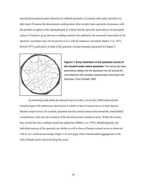
Figure1-7. Early illustration <strong>of</strong> the spectacle nerves <strong>of</strong><br />
the colubrid snake Natrix tessellata. <strong>The</strong> nerves are seen<br />
penetrating radially into the spectacle from all around the<br />
circumference with complex anastomoses occurring in the<br />
branches. From Crevatin 1904.<br />
An interesting aside about the spectacle nerves is that, as Crevatin (1904) observed and<br />
remarked upon with enthusiasm, their layout is similar to that <strong>of</strong> corneal nerves in other species.<br />
Human corneal nerves, for example, penetrate into the corneal stroma from around the corneolimbal<br />
circumference with only the exception <strong>of</strong> the dorsalmost and ventralmost areas. Within the stroma,<br />
they extend fine nerve endings toward the epithelium (Müller et al. 1997). Morphologically, the<br />
individual neurons <strong>of</strong> the spectacle are similar as well to those <strong>of</strong> human corneal nerves as observed<br />
with in vivo confocal microscopy (Figure 1-8, next page) where mitochondrial aggregations in the<br />
form <strong>of</strong> beads can be observed along the axons.<br />
13