Chapter 1, The Reptilian Spectacle - UWSpace - University of ...
Chapter 1, The Reptilian Spectacle - UWSpace - University of ...
Chapter 1, The Reptilian Spectacle - UWSpace - University of ...

You also want an ePaper? Increase the reach of your titles
YUMPU automatically turns print PDFs into web optimized ePapers that Google loves.
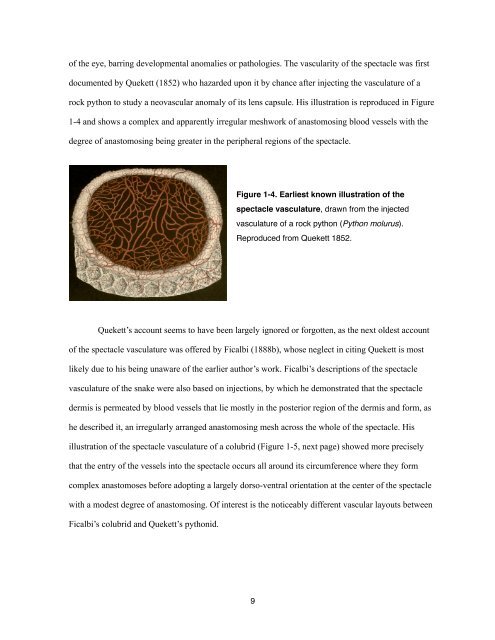
<strong>of</strong> the eye, barring developmental anomalies or pathologies. <strong>The</strong> vascularity <strong>of</strong> the spectacle was first<br />
documented by Quekett (1852) who hazarded upon it by chance after injecting the vasculature <strong>of</strong> a<br />
rock python to study a neovascular anomaly <strong>of</strong> its lens capsule. His illustration is reproduced in Figure<br />
1-4 and shows a complex and apparently irregular meshwork <strong>of</strong> anastomosing blood vessels with the<br />
degree <strong>of</strong> anastomosing being greater in the peripheral regions <strong>of</strong> the spectacle.<br />
Figure 1-4. Earliest known illustration <strong>of</strong> the<br />
spectacle vasculature, drawn from the injected<br />
vasculature <strong>of</strong> a rock python (Python molurus).<br />
Reproduced from Quekett 1852.<br />
Quekett’s account seems to have been largely ignored or forgotten, as the next oldest account<br />
<strong>of</strong> the spectacle vasculature was <strong>of</strong>fered by Ficalbi (1888b), whose neglect in citing Quekett is most<br />
likely due to his being unaware <strong>of</strong> the earlier author’s work. Ficalbi’s descriptions <strong>of</strong> the spectacle<br />
vasculature <strong>of</strong> the snake were also based on injections, by which he demonstrated that the spectacle<br />
dermis is permeated by blood vessels that lie mostly in the posterior region <strong>of</strong> the dermis and form, as<br />
he described it, an irregularly arranged anastomosing mesh across the whole <strong>of</strong> the spectacle. His<br />
illustration <strong>of</strong> the spectacle vasculature <strong>of</strong> a colubrid (Figure 1-5, next page) showed more precisely<br />
that the entry <strong>of</strong> the vessels into the spectacle occurs all around its circumference where they form<br />
complex anastomoses before adopting a largely dorso-ventral orientation at the center <strong>of</strong> the spectacle<br />
with a modest degree <strong>of</strong> anastomosing. Of interest is the noticeably different vascular layouts between<br />
Ficalbi’s colubrid and Quekett’s pythonid.<br />
9