BIOLOGY - microscopia.info
BIOLOGY - microscopia.info
BIOLOGY - microscopia.info
Create successful ePaper yourself
Turn your PDF publications into a flip-book with our unique Google optimized e-Paper software.
98<br />
Overhead Transparency Atlases<br />
fibers, t.s. Axons and myelin sheaths - Various shapes of human neurons - Diagram of a neuron - Various neurons from<br />
human nervous system - Medullated nerve fibers, showing Ranvier’s nodes - The nervous systems - The evolution of<br />
the nervous system in worms - The nervous system of the earthworm - Concentration of ganglia in insects - The head<br />
of a locust l.s. - Position of the brain - The nervous system in arthropods: lobster, crab, spider, scorpion - The nervous<br />
system of a freshwater mussel, a snail and a starfish - Embryonic development of the spinal cord in frog and human -<br />
Human vertebra. Superior and lateral view of three vertebrae with intervertebral discs - Brains of vertebrates (shark,<br />
bony fish, amphibian, reptile, bird, mammal), dorsal views and sagittal sections - Human central nervous system, lateral<br />
view. Position of the dura sac in the spinal canal - Human spinal cord in the spinal canal, lateral view. Opened dural sac,<br />
surface view with segments - Human spinal cord and medulla oblongata. Lateral, dorsal and ventral view with spinal<br />
nerves - Comparison of the masses of brain and spinal cord in Branchiostoma, frog, rabbit, cat, ape, human - Cranial<br />
nerves of frog and sheep - Human brain, ventral view with cranial nerves - Proportion between brain and head in<br />
vertebrates and in mammals - The human spinal cord - Position of the spinal cord in the spinal canal, transection -<br />
Spinal cord of mammal, t.s. silver stained, photomicrograph - Portion of the spinal cord with roots, ganglia, and branches<br />
of spinal nerves, three-dimensional diagram - Simple reflex arc - Tactile corpuscle, spinal cord, motor end plate on<br />
muscle fiber - Polio: syndrome of the ventral gray matter - Tabes, tertiary syphilis: syndrome of the dorsal white matter<br />
- Sclerosis of the pyramidal tracts - Complete section of the spinal cord: Paraplegia - The human brain and the<br />
transmission of <strong>info</strong>rmation - The human brain, lateral view - Sagittal section of the human brain, view on the right<br />
half showing cut surfaces - Frontal section of human brain - The hierarchic structure of the brain, archipallium and<br />
neopallium - Electrotonic or resting potential and action potential - Receptors receive various types of sensory input and<br />
transduce them into action potentials of equal magnitude - Intensity of stimulus is reported by impulse frequency -<br />
Propagation of action potential along unmyelinated axon - Fine structure of a Ranvier’s node (after Krstic) - Nerve cell<br />
body from the cerebrum with dendrites, axon, and synapses - Exciting and inhibiting synapses, their location and<br />
structure - Synapsis, spatial picture - Synaptic transmission, diagram - Brain stem, ventral and dorsal view - The blood<br />
supply of the brain, ventral and lateral view - Lesion caused by diving accident - Lesion caused by hemorrhage (stroke)<br />
- Cerebral cortex, t.s. silver stained to show the pyramidal cells and their connections - The lobes and areas of the left<br />
cerebral hemisphere - Areas and tracts of the cerebrum, diagram - Views of the cerebellum from various sides, and<br />
sagittal section - Fine structure of the cerebellar cortex, neuronal connections, diagram - Tracts connecting the cerebrum<br />
with the cerebellum - The autonomic nervous system - Effect of atropine on one eye, both eyes exposed to<br />
equal incidence of light - Innervation of the iris muscles. Antagonism of sympathetic and parasympathetic nervous<br />
system - Antagonistic effect of the sympathetic and parasympathetic system on glands and involuntary muscle - The<br />
location of the spinal cord, spinal nerves, sympathetic trunk, and sympathetic ganglion II - Transmitter and inhibiting<br />
substances of synapses and motor end plates in the somatic, sympathetic, and parasympathetic nervous system -<br />
Typical courses of sensory and motor tracts of the autonomic nervous system - Regulation of the body temperature -<br />
Location of the receptors and controlling centers in the body, negative feedback system<br />
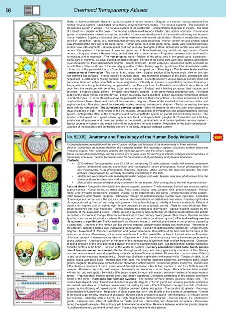
No. 8203E Anatomy and Physiology of the Human Body. Volume III<br />
A comprehensive presentation of the construction, biology and function of the human body in three volumes.<br />
Volume I comprises the human skeleton, the muscular system, the respiratory organs, circulatory system, blood and<br />
lymphatic organs, heart and blood vessels, the digestive system, and the urinary organs.<br />
These atlases of human biology and life science are of great value for teaching in schools, colleges and universities, in<br />
the training of nurses, medical technicians and for the students of physiotherapy and physical education.<br />
Contents:<br />
• 27 Overhead-Transparencies, size 22 x 28 cm, comprising 75 color pictures, mostly with several component<br />
figures (anatomical pictures, photomicro- and macrographs, nature photographs, human photographs, electron<br />
micrographs, X-ray photographs, drawings, diagrams, tables, scenes, test data and results). The color<br />
pictures were prepared by university illustrators specializing in this field.<br />
• Sketch and work-sheets with semidiagrammatic designs and texts. Teacher may take photocopies from the<br />
sheets and use for classroom work and tests.<br />
• Manual with depictured explanatory comments for the teacher. All in strong plastic file with ring-mechanism.<br />
Eye and vision - Range of visible light in the electromagnetic spectrum - The human eye. Eyeball, eye muscles, eyelid,<br />
sagittal section - Human retina, t.s. detail view. Rods, cones, bipolar cells, ganglion cells, photomicrograph - Human<br />
retina. Chief synaptic connections, diagram - Retina, t.s. for detail of rods and cones - Orbital muscles of the eyeball -<br />
Optic pathways, optic chiasm, diagram - Retina seen through the ophthalmoscope. Central fovea, optic disc - Formation<br />
of an image in a normal eye - The eye as a camera - Accommodation for distant and near vision - Pupillary light reflex<br />
- Image produced by “normal” and astigmatic glasses - Eye with pathological turbidity of the lens (cataract) - Defects of<br />
vision: short-sighted and far-sighted eye - Image produced by an astigmatic cornea - Tests for color-blindness. Redgreen<br />
deficiency and blue weakness - Optical illusions by ambiguous <strong>info</strong>rmation - Optical illusions caused by the<br />
influence of the surrounding areas - Optical illusions caused by non-conformity of rational interpretation and optical<br />
perception - Trichromatic triangle. Different combinations of three primary colors give all other colors - Spectral sensitivity<br />
of rods and cones (dominator system), three pigment color vision (modulator system) - Ear and auditory mechanism,<br />
sense of equilibrium - The formation of sound waves. Areas of rarefaction and areas of compression caused by<br />
a tuning fork. - Anatomy of the human ear. Ear concha, external auditory canal, middle ear, internal ear - Movement of<br />
the eardrum, auditory ossicles, oval window and round window - Position of epithelia of the internal ear - Organ of Corti,<br />
diagram - Movement of Reissner’s membrane and basilar membrane. Stimulation of the hair cells by the hairs in the<br />
tectorial membrane - Broadening of the basilar membrane from the base of the cochlea to the helicotrema - Formation<br />
of damped waves in the membranous labyrinth - Displacement of the membranous labyrinth by the waves generated by<br />
sound vibrations - Amplitude pattern of vibration of the membranous labyrinth for high and low frequencies - Detection<br />
of sound direction by the time difference between the entry of sound into the ears - Diagram of main auditory pathways.<br />
Acoustic centers in the brain - Function of the vestibular system - Sensory perception: Smell, taste, touch, perception<br />
of temperature and movement - Section through nasal cavity and pharyngeal cavity - Location of the olfactory<br />
mucous membrane and respiratory pathway - Nasal conchae of human and deer. Microsmates, macrosmates - Olfactory<br />
and respiratory mucous membrane t.s. - Detail view of olfactory epithelium with sensory cilia - Tongue of rabbit, t.s. of<br />
papilla foliata with taste buds - Human skin from palm, v.s. showing cornified epidermis, germinative zone, sweat<br />
glands, diagram - Human scalp, vertical section showing l.s. of hair follicles, sebaceous glands, epidermis - Human skin<br />
with cutaneous receptors of touch, pressure and thermal sensation - Tactile hair, median l.s. and t.s. - Ruffini’s warmth<br />
receptor - Krause’s corpuscle, cold receptor - Meissner’s corpuscle from human finger - Back of human hand marked<br />
with warmth and cold spots - Sensitivity differences caused by touch-stimulation: excitation nearby or far away, weak or<br />
strong - Proprioceptors: muscle spindle and Golgi tendon apparatus. Conscious awareness of the position and movements<br />
of the joints - Hormones and hormone systems - The human hormone glands, position, shape, size - The<br />
human thyroid gland, situs - Exocrine and endocrine glands, diagram - Thyroid gland, sec. showing glandular epithelium<br />
and colloid - Acceleration of tadpole development caused by thyroxin - Effect of thyroxin therapy on a child - Cretinism<br />
caused by insufficiency of thyroid gland - Relation between iodine and goiter - The parathyroid glands - Pancreas<br />
showing islets of Langerhans - Regulation of blood sugar level by A- and B-cells of the islands of Langerhans - Control<br />
of the blood sugar level by insulin and glucagon - Human kidney and adrenal gland - Adrenal gland, t.s. through cortex<br />
and medulla - Interstitial cells of Leydig, t.s., high magnification photomicrograph - Corpus luteum, t.s., photomicrograph<br />
- Castrated fowl, effect of castration on rooster and hen - Secondary sex characters in humans - Processes<br />
during the menstrual cycle - The antibaby pill, hormonal contraception - Relations between endocrine glands, diagram<br />
- Location of pituitary gland and pineal body - Thymus of juvenile and adult person


