

BIOLOGY - microscopia.info
BIOLOGY - microscopia.info
BIOLOGY - microscopia.info

Create successful ePaper yourself
Turn your PDF publications into a flip-book with our unique Google optimized e-Paper software.
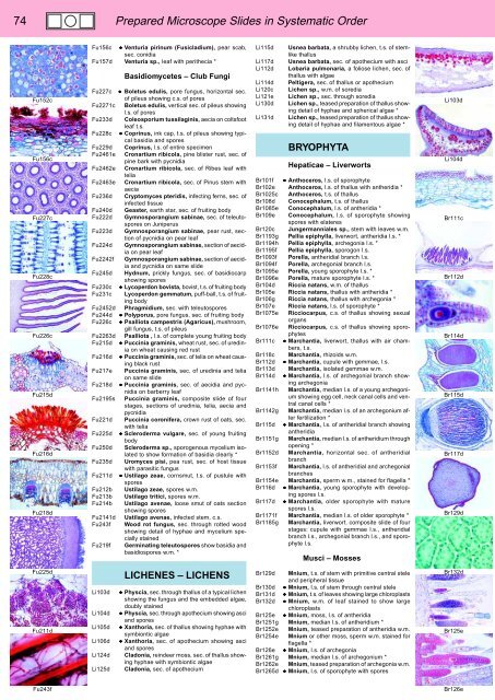
74<br />
Prepared Microscope Slides in Systematic Order<br />
Fu152c<br />
Fu156c<br />
Fu227c<br />
Fu228c<br />
Fu226c<br />
Fu215d<br />
Fu216d<br />
Fu218d<br />
Fu156c • Venturia pirinum (Fusicladium), pear scab,<br />
sec. conidia<br />
Fu157d Venturia sp., leaf with perithecia *<br />
Basidiomycetes – Club Fungi<br />
Fu227c • Boletus edulis, pore fungus, horizontal sec.<br />
of pileus showing c.s. of pores<br />
Fu2271c Boletus edulis, vertical sec. of pileus showing<br />
l.s. of pores<br />
Fu233d Coleosporium tussilaginis, aecia on coltsfoot<br />
leaf t.s.<br />
Fu228c • Coprinus, ink cap, t.s. of pileus showing typical<br />
basidia and spores<br />
Fu229d Coprinus, l.s. of entire specimen<br />
Fu2461e Cronartium ribicola, pine blister rust, sec. of<br />
pine bark with pycnidia<br />
Fu2462e Cronartium ribicola, sec. of Ribes leaf with<br />
telia<br />
Fu2463e Cronartium ribicola, sec. of Pinus stem with<br />
aecia<br />
Fu236d Cryptomyces pteridis, infecting ferns, sec. of<br />
infected tissue<br />
Fu240d Geaster, earth star, sec. of fruiting body<br />
Fu222d Gymnosporangium sabinae, sec. of teleutospores<br />
on Juniperus<br />
Fu223d Gymnosporangium sabinae, pear rust, section<br />
of pycnidia on pear leaf<br />
Fu224d Gymnosporangium sabinae, section of aecidia<br />
on pear leaf<br />
Fu2242f Gymnosporangium sabinae, section of aecidia<br />
and pycnidia on same slide<br />
Fu245d Hydnum, prickly fungus, sec. of basidiocarp<br />
showing spores<br />
Fu230c • Lycoperdon bovista, bovist, t.s. of fruiting body<br />
Fu231c Lycoperdon gemmatum, puff-ball, t.s. of fruiting<br />
body<br />
Fu2452d Phragmidium, sec. with teleutospores<br />
Fu244d • Polyporus, pore fungus, sec. of fruiting body<br />
Fu226c • Psalliota campestris (Agaricus), mushroom,<br />
gill fungus, t.s. of pileus<br />
Fu2263d Psalliota , l.s. of complete young fruiting body<br />
Fu215d • Puccinia graminis, wheat rust, sec. of uredinia<br />
on wheat causing red rust<br />
Fu216d • Puccinia graminis, sec. of telia on wheat causing<br />
black rust<br />
Fu217e Puccinia graminis, sec. of uredinia and telia<br />
on same slide<br />
Fu218d • Puccinia graminis, sec. of aecidia and pycnidia<br />
on barberry leaf<br />
Fu2195s Puccinia graminis, composite slide of four<br />
stages, sections of uredinia, telia, aecia and<br />
pycnidia<br />
Fu221d Puccinia coronifera, crown rust of oats, sec.<br />
with telia<br />
Fu225d • Scleroderma vulgare, sec. of young fruiting<br />
body<br />
Fu250d Scleroderma sp., sporogenous mycelium isolated<br />
to show formation of basidia clearly *<br />
Fu235d Uromyces pisi, pea rust, sec. of host tissue<br />
with parasitic fungus<br />
Fu211d • Ustilago zeae, cornsmut, t.s. of pustule with<br />
spores<br />
Fu212b Ustilago zeae, spores w.m.<br />
Fu213b Ustilago tritici, spores w.m.<br />
Fu214b Ustilago avenae, loose smut of oats section<br />
showing spores<br />
Fu2141d Ustilago avenae, infected stem, c.s.<br />
Fu243f Wood rot fungus, sec. through rotted wood<br />
showing detail of hyphae and mycelium specially<br />
stained<br />
Fu219f Germinating teleutospores show basidia and<br />
basidiospores w.m. *<br />
Li115d<br />
Li117d<br />
Li112d<br />
Li114d<br />
Li120c<br />
Li121e<br />
Li130d<br />
Li131d<br />
Usnea barbata, a shrubby lichen, t.s. of stemlike<br />
thallus<br />
Usnea barbata, sec. of apothecium with asci<br />
Lobaria pulmonaria, a foliose lichen, sec. of<br />
thallus with algae<br />
Peltigera, sec. of thallus or apothecium<br />
Lichen sp., w.m. of soredia<br />
Lichen sp., sec. through soredia<br />
Lichen sp., teased preparation of thallus showing<br />
detail of hyphae and spherical algae *<br />
Lichen sp., teased preparation of thallus showing<br />
detail of hyphae and filamentous algae *<br />
BRYOPHYTA<br />
Hepaticae – Liverworts<br />
Br101f • Anthoceros, l.s. of sporophyte<br />
Br102e Anthoceros, l.s. of thallus with antheridia *<br />
Br1025c Anthoceros, t.s. of thallus<br />
Br108d Conocephalum, t.s. of thallus<br />
Br1085e Conocephalum, l.s. of antheridia *<br />
Br109e Conocephalum, l.s. of sporophyte showing<br />
spores with elateres<br />
Br120c Jungermanniales sp., stem with leaves w.m.<br />
Br1193g Pellia epiphylla, liverwort, antheridia l.s. *<br />
Br1194h Pellia epiphylla, archegonia l.s. *<br />
Br1195f Pellia epiphylla, sporogon l.s.<br />
Br1093f Porella, antheridial branch l.s.<br />
Br1094f Porella, archegonial branch l.s.<br />
Br1095e Porella, young sporophyte l.s. *<br />
Br1096e Porella, mature sporophyte l.s. *<br />
Br104d Riccia natans, w.m. of thallus<br />
Br105e Riccia natans, thallus with antheridia *<br />
Br106g Riccia natans, thallus with archegonia *<br />
Br107e Riccia natans, l.s. of sporophyte *<br />
Br1075e Ricciocarpus, c.s. of thallus showing sexual<br />
organs<br />
Br1076e Ricciocarpus, c.s. of thallus showing sporophytes<br />
Br111c • Marchantia, liverwort, thallus with air chambers,<br />
t.s.<br />
Br118c Marchantia, rhizoids w.m.<br />
Br112d • Marchantia, cupule with gemmae, l.s.<br />
Br113d Marchantia, isolated gemmae w.m.<br />
Br114d • Marchantia, l.s. of archegonial branch showing<br />
archegonia<br />
Br1141h Marchantia, median l.s. of a young archegonium<br />
showing egg cell, neck canal cells and ventral<br />
canal cells *<br />
Br1142g Marchantia, median l.s. of an archegonium after<br />
fertilization *<br />
Br115d • Marchantia, l.s. of antheridial branch showing<br />
antheridia<br />
Br1151g Marchantia, median l.s. of antheridium through<br />
opening *<br />
Br1152d Marchantia, horizontal sec. of antheridial<br />
branch<br />
Br1153f Marchantia, l.s. of antheridial and archegonial<br />
branches<br />
Br1154e Marchantia, sperm w.m., stained for flagella *<br />
Br116d • Marchantia, young sporophyte with developing<br />
spores l.s.<br />
Br117d • Marchantia, older sporophyte with mature<br />
spores l.s.<br />
Br1171f Marchantia, median l.s. of older sporophyte *<br />
Br1185g Marchantia, liverwort. composite slide of four<br />
stages: cupule with gemmae l.s., antheridial<br />
branch l.s., archegonial branch l.s., and sporophyte<br />
l.s.<br />
Musci – Mosses<br />
Li103d<br />
Li104d<br />
Br111c<br />
Br112d<br />
Br114d<br />
Br115d<br />
Br117d<br />
Br129d<br />
Fu225d<br />
Fu211d<br />
Li103d<br />
Li104d<br />
Li105d<br />
Li106d<br />
Li124d<br />
Li125d<br />
LICHENES – LICHENS<br />
• Physcia, sec. through thallus of a typical lichen<br />
showing the fungus and the embedded algae,<br />
doubly stained<br />
• Physcia, sec. through apothecium showing asci<br />
and spores<br />
• Xanthoria, sec. of thallus showing hyphae with<br />
symbiontic algae<br />
• Xanthoria, sec. of apothecium showing asci<br />
and spores<br />
Cladonia, reindeer moss, sec. of thallus showing<br />
hyphae with symbiontic algae<br />
Cladonia, sec. of apothecium<br />
Br129d Mnium, t.s. of stem with primitive central stele<br />
and peripheral tissue<br />
Br130d • Mnium, l.s. of stem through central stele<br />
Br131d • Mnium, t.s. of leaves showing large chloroplasts<br />
Br132d • Mnium, w.m. of leaf stained to show large<br />
chloroplasts<br />
Br125e • Mnium, moss, l.s. of antheridia<br />
Br1251g Mnium, median l.s. of antheridium *<br />
Br1252e Mnium, teased preparation of antheridia w.m.<br />
Br1254e Mnium or other moss, sperm w.m. stained for<br />
flagella *<br />
Br126e • Mnium, l.s. of archegonia<br />
Br1261g Mnium, median l.s. of archegonium *<br />
Br1262e Mnium, teased preparation of archegonia w.m.<br />
Br1265d • Mnium, l.s. of sporophyte with spores<br />
Br132d<br />
Br125e<br />
Fu243f<br />
Br126e


