

BIOLOGY - microscopia.info
BIOLOGY - microscopia.info
BIOLOGY - microscopia.info

You also want an ePaper? Increase the reach of your titles
YUMPU automatically turns print PDFs into web optimized ePapers that Google loves.
124<br />
Overhead Transparency Atlases<br />
Mosses: - Marchantia, liverwort, cupule with gemmae, l.s. - Marchantia, liverwort, l.s. of archegonial branch showing<br />
archegonia - Marchantia, liverwort, l.s. of antheridial branch showing antheridia - Marchantia, liverwort, young sporophyte<br />
with developing spores l.s. - Liverwort (Marchantia) life cycle, all stages of development, color graphic design -<br />
Mnium, moss, t.s. of stem with primitive central stele - Mnium, moss, w.m. of leaf stained to show large chloroplasts -<br />
Mnium, moss, l.s. of antheridia - Mnium, moss, l.s. of archegonia - Moss (Mnium) life cycle, all stages of development,<br />
color graphic design - Mnium, moss, protonema w.m. - Polytrichum, moss, t.s. of leaves showing photosynthetic lamellae<br />
- Polytrichum, t.s. of leaf, color graphic design - Polytrichum, moss, t.s. of stem showing primitive vascular bundle -<br />
Polytrichum, moss, l.s. of sporophyte with spores - Sphagnum, peat moss, w.m. of leaf showing chlorophyll bearing and<br />
hyaline cells.<br />
Ferns and Fern Allies: - Psilotum, primitive fern, t.s. of stem showing exarch protostele - Psilotum, t.s. of three-lobed<br />
sporangium - Lycopodium, club moss, t.s. of stem showing actinostele - Lycopodium, t.s. of mature sporophyll showing<br />
isospores - Lycopodium, anatomy and life-cycle, color graphic design - Equisetum, horsetail, rhizome t.s. - Equisetum,<br />
mature strobilus l.s. - Equisetum, horsetail, life cycle, all stages of development, color graphic design - Equisetum, w.m.<br />
of spores with elaters - Aspidium, (Dryopteris), fern, rhizome t.s. - Aspidium, leaves with l.s. of sori - Aspidium, isolated<br />
sporangia and spores w.m. - Polypodium, leaf with sori and sporangia w.m. - Osmunda, royal fern, rhizome with ectophloic<br />
siphonostele t.s. - Fern prothallium, selected to show antheridia and archegonia w.m. - Fern prothallium, l.s. of<br />
antheridium with spermatozoids - Fern prothallium, l.s. of archegonium with egg cell - Fern life cycle, all stages of<br />
development in 19 pictures, color graphic design.<br />
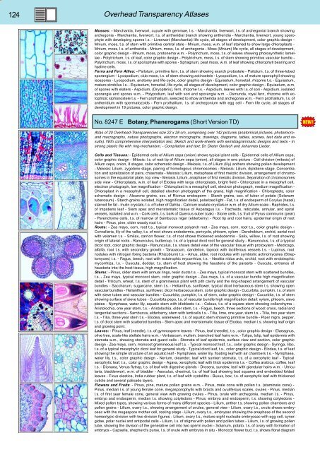
No. 8247 E Botany, Phanerogams (Short Version TD)<br />
Atlas of 20 Overhead-Transparencies size 22 x 28 cm, comprising over 142 pictures (anatomical pictures, photomicroand<br />
macrographs, nature photographs, electron micrographs, drawings, diagrams, tables, scenes, test data and results).<br />
With comprehensive interpretation text. Sketch and work-sheets with semidiagrammatic designs and texts - In<br />
strong plastic file with ring-mechanism. - Compilation and text: Dr. Dieter Gerlach and Johannes Lieder.<br />
Cells and Tissues: - Epidermal cells of Allium cepa (onion) shows typical plant cells - Epidermal cells of Allium cepa,<br />
color graphic design - Mitosis: l.s. of root tip of Allium cepa (onion), all stages in one picture - Cell division (mitosis) of<br />
Allium cepa, onion, 8 stages, color schematic design - Meiosis, t.s. of Lilium (lily) anthers showing pollen development<br />
- Meiosis: Lilium, zygotene stage, pairing of homologous chromosomes - Meiosis: Lilium, diplotene stage. Concentration<br />
and spiralization of pairs, chiasmata - Meiosis: Lilium, metaphase of first meiotic division, arrangement of chromosomes<br />
in the equatorial plate, top view - Meiosis: Lilium, anaphase of first meiotic division. Separation of chromosomes<br />
side view - Chloroplasts, w.m. of leaf of Elodea with large chloroplasts, bright field - Chloroplast in a mesophyll cell,<br />
electron photograph, low magnification - Chloroplast in a mesophyll cell, electron photograph, medium magnification -<br />
Chloroplast in a mesophyll cell, detailed electron photograph of the grana, high magnification - Chloroplasts, color<br />
schematic design - Aleurone grains, sec. of Ricinus endosperm - Starch grains, sec. of tuber of potato (Solanum<br />
tuberosum) - Starch grains isolated, high magnification detail, polarized light - Fat, t.s. of endosperm of Corylus (hazel)<br />
stained for fat - Inulin crystals, t.s. of tuber of Dahlia - Calcium oxalate crystals in w.m. of dry Allium scale - Raphides, t.s.<br />
of Impatiens leaf - Stem apex and meristematic tissue of Asparagus l.s. - Tracheids, reticulate, annular, and spiral<br />
vessels, isolated and w.m. - Cork cells, t.s. bark of Quercus suber (oak) - Stone cells, t.s. fruit of Pyrus communis (pear)<br />
- Parenchyme cells, t.s. of marrow of Sambucus niger (elderberry) - Root tip and root hairs, epidermal origin of root<br />
hairs - Pinus, pine, older woody root t.s.<br />
Roots: - Zea mays, corn, root t.s., typical monocot polyarch root - Zea mays, corn, root t.s., color graphic design -<br />
Convallaria, lily of the valley, t.s. of root shows endodermis, pericycle, phloem, xylem - Dendrobium, orchid, aerial root<br />
with velamen t.s. - Smilax, carrion flower, t.s. of root shows thickened endodermis - Salix, willow, l.s. of root showing<br />
origin of lateral roots - Ranunculus, buttercup, t.s. of a typical dicot root for general study - Ranunculus, t.s. of a typical<br />
dicot root, color graphic design - Ranunculus, t.s. shows detail view of the vascular tissue with protoxylem - Medicago,<br />
alfalfa, root t.s. with secondary growth - Taraxacum, dandelion, taproot with lactiferous vessels t.s. - Lupinus, root<br />
nodules with nitrogen fixing bacteria (Rhizobium) t.s. - Alnus, alder, root nodules with symbiotic actinomycetes (Streptomyces)<br />
t.s. - Fagus, beech, root with ectotrophic mycorrhiza, t.s. - Neottia nidus avis, orchid, root with endotrophic<br />
mycorrhiza, l.s. - Cuscuta, dodder, t.s. stem of host showing the haustoria of the parasite - Cuscuta, entrance of<br />
haustoria into the host tissue, high magnification.<br />
Stems: - Pinus, older stem with annual rings, resin ducts t.s. - Zea mays, typical monocot stem with scattered bundles,<br />
t.s. - Zea mays, typical monocot stem, color graphic design - Zea mays, t.s. of a vascular bundle high magnification<br />
detail - Triticum, wheat, t.s. stem of a gramineous plant with pith cavity and the ring-shaped arrangement of vascular<br />
bundles - Saccharum, sugarcane, stem t.s. - Helianthus, sunflower, typical dicot herbaceous stem t.s. showing open<br />
vascular bundles - Helianthus, sunflower, dicot herbaceous stem, color graphic design - Cucurbita, pumpkin, l.s. of stem<br />
with sieve tubes and vascular bundles - Cucurbita, pumpkin, l.s. of stem, color graphic design - Cucurbita, t.s. of stem<br />
showing surface of sieve tubes - Cucurbita pepo, t.s. of vascular bundle high magnification detail: xylem, phloem, sieve<br />
plates - Nymphaea, water lily, aquatic stem with idioblasts t.s. - Coleus, t.s. of a square stem showing collenchyma -<br />
Aristolochia, one year stem, t.s. - Aristolochia, older stem, t.s. - Fagus, beech, three sections of wood: cross, radial and<br />
tangential sections - Sambucus, elderberry, stem with lenticells t.s. - Tilia, lime, one year, stem t.s. - Tilia, two year stem<br />
t.s. - Tilia, three year stem t.s. - Elodea, waterweed, t.s. of aquatic stem showing primitive bundle - Piper nigra, pepper,<br />
t.s. of dicot stem with scattered bundles - Stem apex and meristematic tissue of Elodea, median l.s. showing leaf origin<br />
and growing point.<br />
Leaves: - Pinus, leaf (needle), t.s. of gymnosperm leaves - Pinus, leaf (needle), t.s., color graphic design - Elaeagnus,<br />
olive tree, scale-like stellate hairs w.m. - Verbascum, mullein, branched leaf hairs w.m. - Tulipa, tulip, leaf epidermis with<br />
stomata w.m., showing stomata and guard cells - Stomata of leaf epidermis, surface view and section, color graphic<br />
design - Zea mays, corn, monocot gramineous leaf t.s. - Typical monocot leaf, t.s., color graphic design - Syringa, lilac,<br />
t.s. of a typical mesophytic dicot leaf for general study - Typical dicot leaf, t.s., color graphic design - Elodea, t.s. of leaf<br />
showing the simple structure of an aquatic leaf - Nymphaea, water lily, floating leaf with air chambers t.s. - Nymphaea,<br />
water lily, t.s., color graphic design - Nerium, oleander, leaf with sunken stomata, t.s. of a xerophytic leaf - Typical<br />
xerophytic leaf, t.s., color graphic design - Agava, xerophytic leaf with thick epidermis t.s. - Coffea arabica, coffee, leaf<br />
t.s. - Dionaea, Venus flytrap, t.s. of leaf with digestive glands - Drosera, sundew, leaf with glandular hairs w.m. - Utricularia,<br />
bladderwort, w.m. of bladder - Aesculus, chestnut, t.s. of leaf bud showing bud squama and embedded folded<br />
leaves - Ficus elastica, India rubber plant, t.s. of leaf with cystoliths - Buxus, box, t.s. of xerophytic leaf with thickened<br />
cuticle and several palisade layers.<br />
Flowers and Fruits: - Pinus, pine, mature pollen grains w.m. - Pinus, male cone with pollen t.s. (staminate cone) -<br />
Pinus, median l.s. of young female cone, megasporophylls with bracts and ovuliferous scales, ovules - Pinus, median<br />
l.s. of first year female cone, general view with growing ovules - Pinus, ovule with archegonia, median l.s. - Pinus,<br />
embryo and endosperm, median l.s. showing cotyledons - Pinus, embryo and endosperm, t.s. showing cotyledons -<br />
Mixed pollen types, showing various forms of many different species - Lilium, anther t.s. showing pollen chambers and<br />
pollen grains - Lilium, ovary t.s., showing arrangement of ovules, general view - Lilium, ovary t.s., ovule shows embryosac<br />
with the megaspore mother cell, resting stage - Lilium, ovary t.s., embryoac showing the anaphase of the second<br />
homeotypic division with two division figures - Lilium, ovary t.s., mature eight nucleate embryosac with egg cell, synergidae,<br />
polar nuclei and antipodal cells - Lilium, l.s. of stigma with pollen and pollen tubes - Lilium, l.s. of growing pollen<br />
tube, showing the division of the generative cell into two sperm nuclei - Solanum, potato, t.s. of ovary with formation of<br />
embryos - Capsella, shepherd’s purse, l.s. of ovule with embryos in situ - Monocot flower bud, t.s. shows floral diagram


