

BIOLOGY - microscopia.info
BIOLOGY - microscopia.info
BIOLOGY - microscopia.info

You also want an ePaper? Increase the reach of your titles
YUMPU automatically turns print PDFs into web optimized ePapers that Google loves.
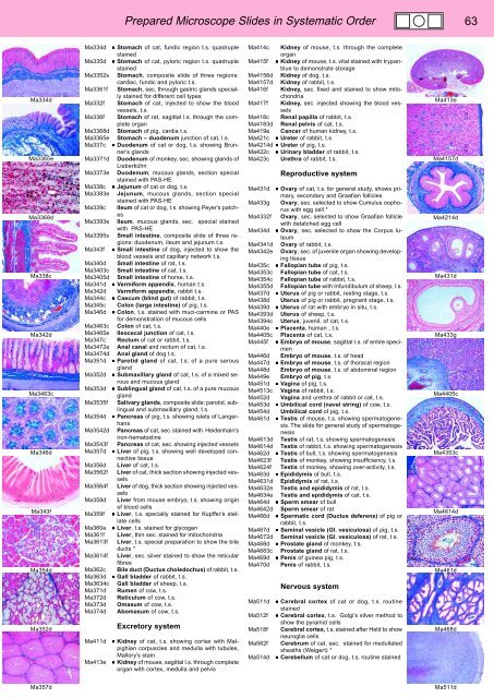
Prepared Microscope Slides in Systematic Order 63<br />
Ma334d<br />
Ma3365e<br />
Ma3368d<br />
Ma338c<br />
Ma342d<br />
Ma3463c<br />
Ma346d<br />
Ma343f<br />
Ma354d<br />
Ma352d<br />
Ma334d • Stomach of cat, fundic region t.s. quadruple<br />
stained<br />
Ma335d • Stomach of cat, pyloric region t.s. quadruple<br />
stained<br />
Ma3352s Stomach, composite slide of three regions:<br />
cardiac, fundic and pyloric t.s.<br />
Ma3361f Stomach, sec. through gastric glands specially<br />
stained for different cell types<br />
Ma332f Stomach of cat, injected to show the blood<br />
vessels, t.s.<br />
Ma336f Stomach of rat, sagittal l.s. through the complete<br />
organ<br />
Ma3368d Stomach of pig, cardia t.s.<br />
Ma3365e Stomach – duodenum junction of cat, l.s.<br />
Ma337c • Duodenum of cat or dog, t.s. showing Brunner’s<br />
glands<br />
Ma3371d Duodenum of monkey, sec. showing glands of<br />
Lieberkühn<br />
Ma3373e Duodenum, mucous glands, section special<br />
stained with PAS-HE<br />
Ma338c • Jejunum of cat or dog, t.s.<br />
Ma3383e Jejunum, mucous glands, section special<br />
stained with PAS-HE<br />
Ma339c Ileum of cat or dog, t.s. showing Peyer’s patches<br />
Ma3393e Ileum, mucous glands, sec. special stained<br />
with PAS-HE<br />
Ma3395s Small intestine, composite slide of three regions:<br />
duodenum, ileum and jejunum t.s.<br />
Ma343f • Small intestine of dog, injected to show the<br />
blood vessels and capillary network t.s.<br />
Ma340d Small intestine of rat, t.s.<br />
Ma3403c Small intestine of cat, t.s.<br />
Ma3405d Small intestine of horse, t.s.<br />
Ma341d • Vermiform appendix, human t.s.<br />
Ma342d Vermiform appendix, rabbit t.s.<br />
Ma344c • Caecum (blind gut) of rabbit, t.s.<br />
Ma345c Colon (large intestine) of pig, t.s.<br />
Ma346d • Colon, t.s. stained with muci-carmine or PAS<br />
for demonstration of mucous cells<br />
Ma3463c Colon of cat, t.s.<br />
Ma3465e Ileocecal junction of cat, l.s.<br />
Ma347c Rectum of cat or rabbit, t.s.<br />
Ma3472e Anal canal and rectum of cat, l.s.<br />
Ma3474d Anal gland of dog t.s.<br />
Ma351d • Parotid gland of cat, t.s. of a pure serous<br />
gland<br />
Ma352d • Submaxillary gland of cat, t.s. of a mixed serous<br />
and mucous gland<br />
Ma353d • Sublingual gland of cat, t.s. of a pure mucous<br />
gland<br />
Ma3535f Salivary glands, composite slide: parotid, sublingual<br />
and submaxillary gland, t.s.<br />
Ma354d • Pancreas of pig, t.s. showing islets of Langerhans<br />
Ma3542d Pancreas of cat, sec. stained with Heidenhain’s<br />
iron-hematoxline<br />
Ma3543f Pancreas of cat, sec. showing injected vessels<br />
Ma357d • Liver of pig, t.s. showing well developed connective<br />
tissue<br />
Ma356d Liver of cat, t.s.<br />
Ma3562f Liver of cat, thick section showing injected vessels<br />
Ma3564f Liver of dog, thick section showing injected vessels<br />
Ma358d Liver from mouse embryo, t.s. showing origin<br />
of blood cells<br />
Ma359f • Liver, t.s. specially stained for Kupffer’s stellate<br />
cells<br />
Ma360e • Liver, t.s. stained for glycogen<br />
Ma361f Liver, thin sec. stained for mitochondria<br />
Ma3613f Liver, t.s. special preparation to show the bile<br />
ducts *<br />
Ma3614f Liver, sec. silver stained to show the reticular<br />
fibres<br />
Ma362c Bile duct (Ductus choledochus) of rabbit, t.s.<br />
Ma363d • Gall bladder of rabbit, t.s.<br />
Ma3634c Gall bladder of sheep, t.s.<br />
Ma371d Rumen of cow, t.s.<br />
Ma372d Reticulum of cow, t.s.<br />
Ma373d Omasum of cow, t.s.<br />
Ma374d Abomasum of cow, t.s.<br />
Ma411d<br />
Ma413e<br />
Excretory system<br />
• Kidney of cat, t.s. showing cortex with Malpighian<br />
corpuscles and medulla with tubules,<br />
Mallory’s stain<br />
• Kidney of mouse, sagittal l.s. through complete<br />
organ with cortex, medulla and pelvis<br />
Ma414c Kidney of mouse, t.s. through the complete<br />
organ<br />
Ma415f • Kidney of mouse, t.s. vital stained with trypanblue<br />
to demonstrate storage<br />
Ma4156d Kidney of dog, t.s.<br />
Ma4157d Kidney of rabbit, t.s.<br />
Ma416f Kidney, sec. fixed and stained to show mitochondria<br />
Ma417f Kidney, sec. injected showing the blood vessels<br />
Ma418c Renal papilla of rabbit, t.s.<br />
Ma4183d Renal pelvis of cat, t.s.<br />
Ma419e Cancer of human kidney, t.s.<br />
Ma421c • Ureter of rabbit, t.s.<br />
Ma4214d • Ureter of pig, t.s.<br />
Ma422c • Urinary bladder of rabbit, t.s.<br />
Ma423c Urethra of rabbit, t.s.<br />
Reproductive system<br />
Ma431d • Ovary of cat, t.s. for general study, shows primary,<br />
secondary and Graafian follicles<br />
Ma433g Ovary, sec. selected to show Cumulus oophorus<br />
with egg cell *<br />
Ma4332f Ovary, sec. selected to show Graafian follicle<br />
with detatched egg cell<br />
Ma434d • Ovary, sec. selected to show the Corpus luteum<br />
Ma4341d Ovary of rabbit, t.s.<br />
Ma4342e Ovary, sec. of juvenile organ showing developing<br />
tissue<br />
Ma435c • Fallopian tube of pig, t.s.<br />
Ma4353c Fallopian tube of cat, t.s.<br />
Ma4354c Fallopian tube of rabbit, t.s.<br />
Ma4355d Fallopian tube with Infundibulum of sheep, l.s.<br />
Ma437d • Uterus of pig or rabbit, resting stage, t.s.<br />
Ma438d Uterus of pig or rabbit, pregnant stage, t.s.<br />
Ma439d • Uterus of rat with embryo in situ, t.s.<br />
Ma4393d Uterus of sheep, t.s.<br />
Ma4394c Uterus, juvenil, of cat, t.s.<br />
Ma440e • Placenta, human , t.s.<br />
Ma4405c Placenta of cat, t.s.<br />
Ma445f • Embryo of mouse, sagittal l.s. of entire specimen<br />
Ma446d Embryo of mouse, t.s. of head<br />
Ma447d • Embryo of mouse, t.s. of thoracal region<br />
Ma448d Embryo of mouse, t.s. of abdominal region<br />
Ma449e Embryo of pig, t.s.<br />
Ma451d • Vagina of pig, t.s.<br />
Ma4513c Vagina of rabbit, t.s.<br />
Ma452d Vagina and urethra of rabbit or cat, t.s.<br />
Ma453d • Umbilical cord (navel string) of cow, t.s.<br />
Ma454d Umbilical cord of pig, t.s.<br />
Ma461d • Testis of mouse, t.s. showing spermatogenesis.<br />
The slide for general study of spermatogenesis<br />
Ma4613d Testis of rat, t.s. showing spermatogenesis<br />
Ma4614d Testis of rabbit, t.s. showing spermatogenesis<br />
Ma462d • Testis of bull, t.s. showing spermatogenesis<br />
Ma4623f Testis of monkey, showing insufficiency, t.s.<br />
Ma4624f Testis of monkey, showing over-activity, t.s.<br />
Ma463d • Epididymis of bull, t.s.<br />
Ma4631d Epididymis of rat, t.s.<br />
Ma4632e Testis and epididymis of rat, t.s.<br />
Ma4634e Testis and epididymis of cat, t.s.<br />
Ma464d • Sperm smear of bull<br />
Ma4642d Sperm smear of rat<br />
Ma466d • Spermatic cord (Ductus deferens) of pig or<br />
rabbit, t.s.<br />
Ma467d • Seminal vesicle (Gl. vesiculosa) of pig, t.s.<br />
Ma4672d Seminal vesicle (Gl. vesiculosa) of rat, t.s.<br />
Ma468d • Prostate gland of monkey, t.s.<br />
Ma4683c Prostate gland of rat, t.s.<br />
Ma469d • Penis of guinea pig, t.s.<br />
Ma470d Penis of rabbit, t.s.<br />
Ma511d<br />
Ma512f<br />
Ma518f<br />
Ma562f<br />
Ma514d<br />
Nervous system<br />
• Cerebral cortex of cat or dog, t.s. routine<br />
stained<br />
• Cerebral cortex, t.s. Golgi’s silver method to<br />
show the pyramid cells<br />
Cerebral cortex, t.s. stained after Held to show<br />
neuroglia cells<br />
Cerebrum of cat, sec. stained for medullated<br />
sheaths (Weigert) *<br />
• Cerebellum of cat or dog, t.s. routine stained<br />
Ma413e<br />
Ma4157d<br />
Ma4214d<br />
Ma431d<br />
Ma433g<br />
Ma4405c<br />
Ma4353c<br />
Ma4614d<br />
Ma461d<br />
Ma468d<br />
Ma357d<br />
Ma511d


