

BIOLOGY - microscopia.info
BIOLOGY - microscopia.info
BIOLOGY - microscopia.info

You also want an ePaper? Increase the reach of your titles
YUMPU automatically turns print PDFs into web optimized ePapers that Google loves.
Overhead Transparency Atlases<br />
115<br />
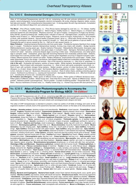
No. 8250 E Environmental Damages (Short Version TH)<br />
Atlas of 18 Overhead-Transparencies size 22 x 28 cm, comprising over 80 color pictures (photomicro- and macrographs,<br />
nature photographs, human photographs, electron micrographs, life cycles, drawings, diagrams, tables, scenes,<br />
test data and results). - With comprehensive interpretation text. - In strong plastic file with ring-mechanism. Compilation<br />
and text: Dr. Karl-Heinrich Meyer B.S. and Johannes Lieder.<br />
The Wood: - Pine (Pinus), healthy leaves, t.s. - Pine (Pinus) leaves damaged by acid rain, t.s. - Fir (Abies), healthy<br />
leaves, t.s. - Fir (Abies), stem tip damaged t.s. - Beech (Fagus), healthy leaves t.s. - Beech (Fagus), t.s. of leaves with<br />
destroyed epidermis and chloroplasts - Rhytisma acerinum, tar spot of maples, consequence of single-crop farming -<br />
Early leaf fall, caused by thawing salt - Healthy lichen, indicator of clean air - Damaged lichen, caused by air pollution -<br />
Healthy wood of beech, t.s. - Wood destroyed by fungus - Polyporus, wood rot fungus, fruiting body t.s. - Root nodules<br />
of Alnus, with symbiotic bacteria - Spruce beetle (Cryphalus picea), larva t.s. - Wood with normal annual rings, t.s. -<br />
Wood with anomalous narrow annual rings caused by drought, t.s. - Bark with larval galleries of spruce beetle, t.s. -<br />
Pineapple-like gall on spruce caused by lice, t.s. - Gall nut on oak caused insects, t.s.<br />
Water Pollution: - Intestinal bacteria (Escherichia coli) from putrid water - Putrefactive bacteria (Spirillum) from sludge<br />
poor in oxygen - Putrefactive bacteria (Sphaerotilus) bacteria, forming long chains with sheaths - Sludge bacteria<br />
(Methanobacterium) causing sewer gas - Sulphur bacteria (Thiocystis) - Wasserbluthe (Microcystis), blue-green alga<br />
“blooming” in stagnant water - Anabaena, blue green algae, in eutrophic water - Spirogyra, filamentous green alga in<br />
nutrient-rich water - Spirulina, corkscrew-shaped algae occurring in bitter seas - Chlamydomonas, one-celled green<br />
alga in eutrophic water - Cladophora, green alga with branching filaments from moderately polluted water - Diatoms,<br />
mixed algae from scarcely polluted water - Euglena, common green flagellates occurring in stagnant eutrophic water -<br />
Ciliates, different species from nutrient-rich water - Rotifers (Rotatoria), small animals from putrid water - Tubifex, fresh<br />
water oligochaete, living in the sludge - Carchesium, bell-shaped stalked ciliate from moderately polluted water - Water<br />
mold (Saprolegnia), harmful to plants and animals - Skin of fish injured by chemicals, t.s. - Skin ulcer of amphibian, t.s.<br />
Life in the soil: - Acidophile soil bacteria, solution of heavy metals - Nitrite bacteria, formatting harmful nitrogenous<br />
substances - Root of beech with ectotrophic mycorrhiza, t.s. - Root of birch with partly endotrophic mycorrhiza, t.s. -<br />
Root of lupin with symbiotic nitrogen fixing bacteria - Netted venation, portion of rotted deciduous leaf - Charlock<br />
(Sinapis), t.s. of stem. Green manure plant - Soil bacteria (Bacillus megatherium), smear - Hyphae of root fungi, t.s. -<br />
Lichen, indicator of clean air - Mushroom (Xerocomus), mycelium - Root of willow (Salix), planting protecting against<br />
erosion - Earthworm (Lumbricus) t.s., causing soil improvement - Springtails (Collembola), w.m. - Mite from forest soil,<br />
w.m. - Constituents of humus soil - Constituents of peaty soil.<br />
Air Pollution and Allergens: - Pollen grains of different kinds of grass - Pollen grains of different deciduous trees -<br />
Pollen grains of different conifers - Mixed house dust - Dust mite from a living room - Spores of different fungi - Wood<br />
powder - Asbestos powder (cancerogenous) - Talcum powder - Crystals of washing-powder - Polyamide fibers - Nylon<br />
fibers - Mucous membrane of human nose, t.s. - Healthy human lung, t.s. - Human lung injured with dust particles, t.s.<br />
No. 8236 E Atlas of Color Photomicrographs to Accompany the<br />
Multimedia-Program for Biology ABCD 7th Edition!<br />
Atlas of 45 OHP Transparencies size 22 x 28 cm, comprising over 252 color photomicrographs according to the 175<br />
Prepared Microscope Slides of the MULTIMEDIA-SYSTEM FOR <strong>BIOLOGY</strong> A, B, C and D (see pages 4 - 10). Detailed<br />
explanatory textbook. Plus new sketch and work-sheets with semidiagrammatic designs and texts.<br />
This atlas of OHP transparencies is intended to present a clear-cut outline of all fields of biology and cover all the<br />
organisms studied in schools. Each of the specimens has been carefully chosen on the basis of its instructional value.<br />
Zoology. - Amoeba proteus, showing nucleus and pseudopodia - Radiolaria, mixed species - Foraminifera, mixed<br />
species - Euglena, flagellate with eyespot - Trypanosoma gambiense, sleeping disease, blood smear - Plasmodium<br />
berghei, malaria parasite, blood smear - Paramecium, nuclei stained - Sycon, marine sponge t.s. - Hydra, w.m.<br />
extended specimen - Hydra, t.s. of body - Obelia hydroid, w.m. of colony - Planaria, typical t.s. - Dicrocoelium<br />
lanceolatum, sheep liver fluke w.m. - Distomum hepaticum (Fasciola), beef liver fluke w.m. - Taenia saginata, tapeworm,<br />
proglottids t.s. - Taenia, tapeworm, w.m. of mature proglottids - Trichinella spiralis, l.s. of skeletal muscle<br />
showing encysted larvae - Ascaris, roundworm, t.s. of female in region of gonads - Lumbricus, earthworm, typical t.s.<br />
back of clitellum - Daphnia and Cyclops, small crustaceans - Araneus, spider, leg with comb w.m. - Araneus, spinneret<br />
w.m. - Dermanyssus gallinae, chicken mite, w.m. - Musca domestica, house fly, head and mouth parts - Musca<br />
domestica, leg with clinging pads - Apis mellifica, honey bee, mouth parts of worker - Apis mellifica, honey bee,<br />
wings - Apis mellifica, hind leg of worker with pollen basket - Apis mellifica, sting and poison sac - Apis mellifica,<br />
head with compound eyes and brain t.s. - Apis mellifica, abdomen of worker t.s. with intestine and nephridia - Periplaneta,<br />
cockroach, chewing mouth parts - Culex pipiens, mosquito, head and piercing-sucking mouth parts of female - Culex<br />
pipiens, mosquito, reduced mouth parts of male - Trachea from insect - Spiracle from insect - Pieris, butterfly, portion<br />
of wing with scales - Ctenocephalus canis, dog flea, w.m. - Cimex lectularius, bed bug, w.m. - Helix pomatia, snail,<br />
hermaphrodite gland (ovotestis), t.s. with developing ova and spermatozoa - Mya arenaria, clam, gill sec. with ciliated<br />
epithelium - Bird feathers, wing or vane and down feathers - Asterias rubens, starfish, arm (ray) t.s. showing tube<br />
feet, digestive gland, ampullae - Branchiostoma lanceolatum (Amphioxus), t.s. of body with gills, liver, and gonads<br />
Histology of Man and Mammals. - Squamous epithelium, isolated cells from human mouth - Ciliated epithelium, in<br />
t.s. of fallopian tube - Fibrous connective tissue of mammal - Tendon of cow, l.s. white fibrous tissue - Adipose<br />
tissue, stained for fat - Hyaline cartilage t.s. - Compact bone, t.s. cells, lamellae, and canaliculi - Striated muscle, l.s.<br />
showing nuclei and striations - Heart muscle, human, l.s. branched fibers with intercalated discs - Smooth (involuntary)<br />
muscle l.s. and t.s. - Lung of cat, t.s. showing alveoli - Human blood smear, red and white corpuscles - Frog<br />
blood smear, nucleated red corpuscles - Artery and vein of mammal, t.s. - Lymph gland of pig, t.s. showing lymphoid<br />
tissue - Thyroid gland of pig, sec. showing colloid - Adrenal gland of cat, t.s. through cortex and medulla - Esophagus<br />
of cat, t.s. - Stomach of cat, t.s. fundic region - Small intestine of cat, t.s. - Large intestine (colon), t.s. stained for<br />
mucous cells - Liver of pig, t.s. - Pancreas of pig, sec. with islets of Langerhans - Kidney of cat, t.s. through cortex and<br />
medulla - Ovary of cat, t.s. with primary, secondary, and Graafian follicles - Testis of mouse, t.s. showing spermatogenesis<br />
- Sperm of bull, smear - Medullated nerve fibers, osmic acid fixed showing Ranvier’s nodes - Motor nerve cells,<br />
smear from spinal cord of cow - Spinal cord of cat, t.s. white and grey matter - Cerebrum, human, t.s. of cortex with<br />
pyramidal cells - Cerebellum of cat, t.s. shows Purkinje cells - Retina of cat, t.s. detail of rods and cones - Tongue of<br />
rabbit, t.s. of papilla foliata with taste buds - Human skin from palm, v.s. sweat glands - Human scalp, l.s. of hair<br />
follicles<br />
Botany, Bacteria and Cryptogams. - Bacteria from mouth, smear Gram stained showing bacilli, cocci, spirilli, spirochaetes<br />
- Bacillus subtilis, hay bacillus, smear with bacilli and spores - Streptococcus lactis, milk souring organisms<br />
- Oscillatoria, a blue green filamentous alga - Nostoc, blue green alga, colonies within gelatinous sheaths - Diatoms,<br />
mixed species - Cladophora, green alga, branched filaments with multinucleate cells - Volvox, with daughter colonies<br />
and sexual stages - Spirogyra, vegetative filaments with spiral chloroplasts - Spirogyra in scalariform conjugation,<br />
formation of zygotes - Desmids (Desmidiaceae), various species - Fucus vesiculosus, brown alga, female conceptacle<br />
with oogonia t.s. - Fucus vesiculosus, male conceptacle with antheridia t.s. - Mucor or Rhizopus, mold, w.m. of


