

BIOLOGY - microscopia.info
BIOLOGY - microscopia.info
BIOLOGY - microscopia.info

Create successful ePaper yourself
Turn your PDF publications into a flip-book with our unique Google optimized e-Paper software.
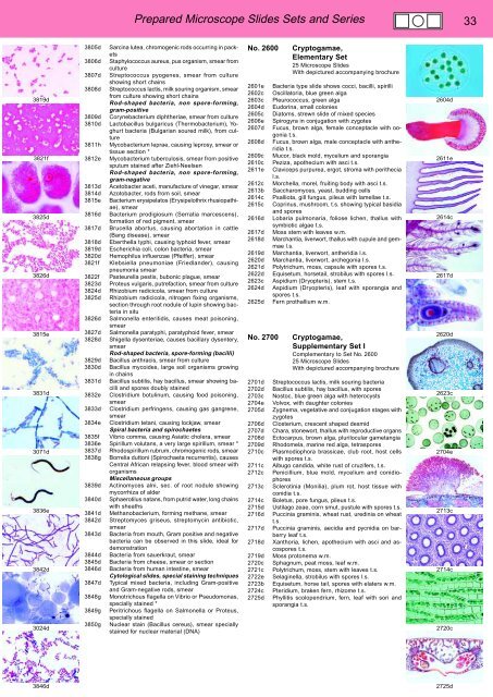
Prepared Microscope Slides Sets and Series 33<br />
3819d<br />
3821f<br />
3825d<br />
3826d<br />
3815e<br />
3831d<br />
3071d<br />
3836e<br />
3842d<br />
3024d<br />
3805d Sarcina lutea, chromogenic rods occurring in packets<br />
3806d Staphylococcus aureus, pus organism, smear from<br />
culture<br />
3807d Streptococcus pyogenes, smear from culture<br />
showing short chains<br />
3808d Streptococcus lactis, milk souring organism, smear<br />
from culture showing short chains<br />
Rod-shaped bacteria, non spore-forming,<br />
gram-positive<br />
3809d Corynebacterium diphtheriae, smear from culture<br />
3810d Lactobacillus bulgaricus (Thermobacterium), Yoghurt<br />
bacteria (Bulgarian soured milk), from culture<br />
3811h Mycobacterium leprae, causing leprosy, smear or<br />
tissue section *<br />
3812e Mycobacterium tuberculosis, smear from positive<br />
sputum stained after Ziehl-Neelsen<br />
Rod-shaped bacteria, non spore-forming,<br />
gram-negative<br />
3813d Acetobacter aceti, manufacture of vinegar, smear<br />
3814d Azotobacter, rods from soil, smear<br />
3815e Bacterium erysipelatos (Erysipelothrix rhusiopathiae),<br />
smear<br />
3816d Bacterium prodigiosum (Serratia marcescens),<br />
formation of red pigment, smear<br />
3817d Brucella abortus, causing abortation in cattle<br />
(Bang disease), smear<br />
3818d Eberthella typhi, causing typhoid fever, smear<br />
3819d Escherichia coli, colon bacteria, smear<br />
3820d Hemophilus influenzae (Pfeiffer), smear<br />
3821f Klebsiella pneumoniae (Friedlander), causing<br />
pneumonia smear<br />
3822f Pasteurella pestis, bubonic plague, smear<br />
3823d Proteus vulgaris, putrefaction, smear from culture<br />
3824d Rhizobium radicicola, smear from culture<br />
3825d Rhizobium radicicola, nitrogen fixing organisms,<br />
section through root nodule of lupin showing bacteria<br />
in situ<br />
3826d Salmonella enteritidis, causes meat poisoning,<br />
smear<br />
3827d Salmonella paratyphi, paratyphoid fever, smear<br />
3828d Shigella dysenteriae, causes bacillary dysentery,<br />
smear<br />
Rod-shaped bacteria, spore-forming (bacilli)<br />
3829d Bacillus anthracis, smear from culture<br />
3830d Bacillus mycoides, large soil organisms growing<br />
in chains<br />
3831d Bacillus subtilis, hay bacillus, smear showing bacilli<br />
and spores doubly stained<br />
3832e Clostridium botulinum, causing food poisoning,<br />
smear<br />
3833d Clostridium perfringens, causing gas gangrene,<br />
smear<br />
3834e Clostridium tetani, causing lockjaw, smear<br />
Spiral bacteria and spirochaetes<br />
3835f Vibrio comma, causing Asiatic cholera, smear<br />
3836e Spirillum volutans, a very large spirillum, smear *<br />
3837d Rhodospirillum rubrum, chromogenic rods, smear<br />
3838g Borrelia duttoni (Spirochaeta recurrentis), causes<br />
Central African relapsing fever, blood smear with<br />
organisms<br />
Miscellaneous groups<br />
3839d Actinomyces alni, sec. of root nodule showing<br />
mycorrhiza of alder<br />
3840d Sphaerotilus natans, from putrid water, long chains<br />
with sheaths<br />
3841d Methanobacterium, forming methane, smear<br />
3842d Streptomyces griseus, streptomycin antibiotic,<br />
smear<br />
3843d Bacteria from mouth, Gram positive and negative<br />
bacteria can be observed in this slide, ideal for<br />
demonstration<br />
3844d Bacteria from sauerkraut, smear<br />
3845d Bacteria from cheese, smear or section<br />
3846d Bacteria from human intestine, smear<br />
Cytological slides, special staining techniques<br />
3847d Typical mixed bacteria, including Gram-positive<br />
and Gram-negative rods, smear<br />
3848g Monotrichous flagella on Vibrio or Pseudomonas,<br />
specially stained *<br />
3849g Peritrichous flagella on Salmonella or Proteus,<br />
specially stained<br />
3850g Nuclear stain (Bacillus cereus), smear specially<br />
stained for nuclear material (DNA)<br />
No. 2600<br />
2601e<br />
2602c<br />
2603c<br />
2604d<br />
2605c<br />
2606e<br />
2607d<br />
2608d<br />
2609c<br />
2610c<br />
2611e<br />
2612c<br />
2613b<br />
2614c<br />
2615c<br />
2616d<br />
2617d<br />
2618d<br />
2619d<br />
2620d<br />
2621d<br />
2622d<br />
2623c<br />
2624d<br />
2625d<br />
No. 2700<br />
2701d<br />
2702d<br />
2703c<br />
2704e<br />
2705d<br />
2706d<br />
2707d<br />
2708d<br />
2709d<br />
2710c<br />
2711c<br />
2712c<br />
2713c<br />
2714c<br />
2715d<br />
2716d<br />
2717d<br />
2718d<br />
2719d<br />
2720c<br />
2721c<br />
2722e<br />
2723b<br />
2724c<br />
2725d<br />
Cryptogamae,<br />
Elementary Set<br />
25 Microscope Slides<br />
With depictured accompanying brochure<br />
Bacteria type slide shows cocci, bacilli, spirilli<br />
Oscillatoria, blue green alga<br />
Pleurococcus, green alga<br />
Eudorina, small colonies<br />
Diatoms, strewn slide of mixed species<br />
Spirogyra in conjugation with zygotes<br />
Fucus, brown alga, female conceptacle with oogonia<br />
t.s.<br />
Fucus, brown alga, male conceptacle with antheridia<br />
t.s.<br />
Mucor, black mold, mycelium and sporangia<br />
Peziza, apothecium with asci t.s.<br />
Claviceps purpurea, ergot, stroma with perithecia<br />
l.s.<br />
Morchella, morel, fruiting body with asci t.s.<br />
Saccharomyces, yeast, budding cells<br />
Psalliota, gill fungus, pileus with lamellae t.s.<br />
Coprinus, mushroom, t.s. showing typical basidia<br />
and spores<br />
Lobaria pulmonaria, foliose lichen, thallus with<br />
symbiotic algae t.s.<br />
Moss stem with leaves w.m.<br />
Marchantia, liverwort, thallus with cupule and gemmae<br />
l.s.<br />
Marchantia, liverwort, antheridia l.s.<br />
Marchantia, liverwort, archegonia l.s.<br />
Polytrichum, moss, capsule with spores t.s.<br />
Equisetum, horsetail, strobilus with spores l.s.<br />
Aspidium (Dryopteris), stem t.s.<br />
Aspidium (Dryopteris), leaf with sporangia and<br />
spores t.s.<br />
Fern prothallium w.m.<br />
Cryptogamae,<br />
Supplementary Set I<br />
Complementary to Set No. 2600<br />
25 Microscope Slides<br />
With depictured accompanying brochure<br />
Streptococcus lactis, milk souring bacteria<br />
Bacillus subtilis, hay bacillus, with spores<br />
Nostoc, blue green alga with heterocysts<br />
Volvox, with daughter colonies<br />
Zygnema, vegetative and conjugation stages with<br />
zygotes<br />
Closterium, crescent shaped desmid<br />
Chara, stonewort, thallus with reproductive organs<br />
Ectocarpus, brown alga, plurilocular gametangia<br />
Rhodomela, marine red alga, tetraspores<br />
Plasmodiophora brassicae, club root, host cells<br />
with spores t.s.<br />
Albugo candida, white rust of cruzifers, t.s.<br />
Penicillium, blue mold, mycelium and conidiophores<br />
Sclerotinia (Monilia), plum rot, host tissue with<br />
conidia t.s.<br />
Boletus, pore fungus, pileus t.s.<br />
Ustilago zeae, corn smut, pustule with spores t.s.<br />
Puccinia graminis, wheat rust, uredinia on wheat<br />
t.s.<br />
Puccinia graminis, aecidia and pycnidia on barberry<br />
leaf t.s.<br />
Xanthoria, lichen, apothecium with asci and ascospores<br />
t.s.<br />
Moss protonema w.m.<br />
Sphagnum, peat moss, leaf w.m.<br />
Polytrichum, moss, stem with leaves t.s.<br />
Selaginella, strobilus with spores l.s.<br />
Equisetum, horse tail, spores with elaters w.m.<br />
Pteridium, braken fern, rhizome t.s.<br />
Phyllitis scolopendrium, fern, leaf with sori and<br />
sporangia t.s.<br />
2604d<br />
2611e<br />
2614c<br />
2617d<br />
2620d<br />
2623c<br />
2704e<br />
2713c<br />
2714c<br />
2720c<br />
3846d<br />
2725d


