

BIOLOGY - microscopia.info
BIOLOGY - microscopia.info
BIOLOGY - microscopia.info

You also want an ePaper? Increase the reach of your titles
YUMPU automatically turns print PDFs into web optimized ePapers that Google loves.
MEDIEN<br />
Multimedia Program Microscopic Biology ABCD<br />
SYSTEM 9<br />
3. Color Atlas of Overhead Projector Transparencies<br />
New 7th Edition 2011<br />
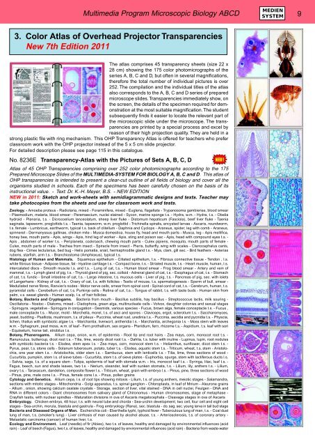
The atlas comprises 45 transparency sheets (size 22 x<br />
28 cm) showing the 175 color photomicrographs of the<br />
series A, B, C and D, but often in several magnifications,<br />
therefore the total number of individual pictures is over<br />
252. The compilation and the individual titles of the atlas<br />
also corresponds to the A, B, C and D series of prepared<br />
microscope slides. Transparencies immediately show, on<br />
the screen, the details of the specimen required for demonstration<br />
at the most suitable magnification. The student<br />
subsequently finds it easier to locate the relevant part of<br />
the microscopic slide under the microscope. The transparencies<br />
are printed by a special process and excel by<br />
reason of their high projection quality. They are held in a<br />
strong plastic file with ring mechanism. This OHP Transparency Atlas is offered for teachers who prefer<br />
classroom work with the OHP projector instead of the 5 x 5 cm slide projector.<br />
For detailed description please see page 115 in this catalogue.<br />
No. 8236E Transparency-Atlas with the Pictures of Sets A, B, C, D<br />
Atlas of 45 OHP Transparencies comprising over 252 color photomicrographs according to the 175<br />
Prepared Microscope Slides of the MULTIMEDIA-SYSTEM FOR <strong>BIOLOGY</strong> A, B, C and D. This atlas of<br />
OHP transparencies is intended to present a clear-cut outline of all fields of biology and cover all the<br />
organisms studied in schools. Each of the specimens has been carefully chosen on the basis of its<br />
instructional value. - Text: Dr. K.-H. Meyer, B.S. - NEW EDITION<br />
NEW in 2011: Sketch and work-sheets with semidiagrammatic designs and texts. Teacher may<br />
take photocopies from the sheets and use for classroom work and tests.<br />
Zoology. - Amoeba proteus - Radiolaria, mixed - Foraminifera, mixed - Euglena, flagellate - Trypanosoma gambiense, blood smear<br />
- Plasmodium, malaria, blood smear - Paramaecium, nuclei stained - Sycon, marine sponge t.s. - Hydra, w.m. - Hydra, t.s. - Obelia<br />
hydroid - Planaria, t.s. - Dicrocoelium lanceolatum, sheep liver fluke - Distomum hepaticum (Fasciola), beef liver fluke - Taenia<br />
saginata, tapeworm, proglottids t.s. - Taenia, tapeworm, w.m. proglottid - Trichinella spiralis, encysted larvae - Ascaris, roundworm,<br />
t.s. female - Lumbricus, earthworm, typical t.s. back of clitellum - Daphnia and Cyclops - Araneus, spider, leg with comb - Araneus,<br />
spinneret - Dermanyssus gallinae, chicken mite - Musca domestica, house fly, head and mouth parts - Musca, leg - Apis mellifica,<br />
honey bee, mouth parts - Apis, wings - Apis, hind leg of worker - Apis, sting and poison sac - Apis, head with compound eyes t.s. -<br />
Apis , abdomen of worker t.s. - Periplaneta, cockroach, chewing mouth parts - Culex pipiens, mosquito, mouth parts of female -<br />
Culex, mouth parts of male - Trachea from insect - Spiracle from insect - Pieris, butterfly, wing with scales - Ctenocephalus canis,<br />
dog flea - Cimex lectularius, bed bug - Helix pomatia, snail, hermaphrodite gland t.s. - Mya, clam, gill sec. - Bird feathers - Asterias<br />
rubens, starfish, arm t.s. - Branchiostoma (Amphioxus), typical t.s.<br />
Histology of Human and Mammals. Squamous epithelium - Ciliated epithelium, t.s. - Fibrous connective tissue - Tendon , l.s.<br />
white fibrous tissue - Adipose tissue, fat - Hyaline cartilage t.s. - Compact bone, t.s. - Striated muscle, l.s. - Heart muscle, human, l.s.<br />
intercalated discs - Smooth muscle l.s. and t.s. - Lung of cat, t.s. - Human blood smear - Frog blood smear - Artery and vein of<br />
mammal, t.s. - Lymph gland of pig, t.s. - Thyroid gland of pig, sec. colloid - Adrenal gland of cat, t.s. - Esophagus of cat, t.s. - Stomach<br />
of cat, t.s. fundic - Small intestine of cat, t.s. - Large intestine, t.s. mucous cells - Liver of pig, t.s. - Pancreas of pig, sec. with islets<br />
of Langerhans - Kidney of cat, t.s. - Ovary of cat, t.s. with follicles - Testis of mouse, t.s. spermatogenesis - Sperm of bull, smear -<br />
Medullated nerve fibres, Ranvier’s nodes - Motor nerve cells, smear from spinal cord - Spinal cord of cat, t.s. - Cerebrum, human, t.s.<br />
pyramidal cells - Cerebellum of cat, t.s. Purkinje cells - Retina of cat, t.s. - Tongue of rabbit, t.s. with taste buds - Human skin from<br />
palm, v.s. sweat glands - Human scalp, l.s. of hair follicles<br />
Botany, Bacteria and Cryptogams. Bacteria from mouth - Bacillus subtilis, hay bacillus - Streptococcus lactis, milk souring -<br />
Oscillatoria - Nostoc - Diatoms, mixed - Cladophora, green alga, multinucleate cells - Volvox, daughter colonies and sexual stages<br />
- Spirogyra, vegetative - Spirogyra in conjugation - Desmids, various species - Fucus, brown alga, female conceptacle t.s. - Fucus ,<br />
male conceptacle t.s. - Mucor, mold - Morchella, morel, t.s. of asci and spores - Claviceps, ergot, sclerotium t.s. - Saccharomyces,<br />
yeast, budding - Psalliota, mushroom, t.s. of pileus - Puccinia, wheat rust, uredinia t.s. - Puccinia, aecidia and pycnidia t.s. - Physcia,<br />
lichen, thallus with symbiotic algae t.s. - Marchantia, liverwort, antheridia l.s. - Marchantia, archegonia l.s. - Moss stem with leaves<br />
w.m. - Sphagnum, peat moss, w.m. of leaf - Fern prothallium, sex organs - Pteridium, fern, rhizome t.s. - Aspidium, t.s. leaf with sori<br />
- Equisetum, horse tail, strobilus l.s.<br />
Botany, Phanerogams. Allium cepa, onion, w.m. of epidermis - Root tip and root hairs - Zea mays, corn, monocot root t.s. -<br />
Ranunculus, buttercup, dicot root t.s. - Tilia, lime, woody dicot root t.s. - Dahlia, t.s. tuber with inuline - Lupinus, lupin, root nodules<br />
with symbiotic bacteria t.s. - Elodea, stem apex l.s. - Zea mays, corn, monocot stem t.s. - Helianthus, sunflower, dicot stem t.s. -<br />
Pyrus, pear, t.s. stone cells - Solanum tuberosum, potato, tuber t.s. - Elodea, aquatic stem t.s. - Triticum, wheat, t.s. stem - Aristolochia,<br />
one year stem t.s. - Aristolochia, older stem t.s. - Sambucus, stem with lenticells t.s. - Tilia, lime, three sections of wood -<br />
Cucurbita, pumpkin, stem l.s. of sieve tubes - Cucurbita, stem t.s. of sieve plates - Euphorbia, spurge, stem with lactiferous ducts l.s.<br />
- Salvia, sage, t.s. of a square stem - Tulipa, epidermis of leaf with stomata w.m. - Iris, monocot leaf t.s. - Syringa, lilac, leaf t.s. -<br />
Fagus, beech, sun and shade leaves, two t.s. - Nerium, oleander, leaf with sunken stomata, t.s. - Lilium, lily, anthers t.s. - Lilium,<br />
ovary t.s. - Taraxacum, dandelion, composite flower l.s. - Triticum, wheat, grain with embryo l.s. - Pinus, pine, three sections of wood<br />
- Pinus, pine, male cone l.s. - Pinus, female cone l.s. - Pinus, pollen grains<br />
Cytology and Genetics. Allium cepa, l.s. of root tips showing mitosis - Lilium, t.s. of young anthers, meiotic stages - Salamandra,<br />
sections with mitotic stages - Mitochondria - Golgi apparatus, t.s. spinal ganglion - Chloroplasts, in leaf of Mnium - Aleurone grains<br />
- Allium , onion, showing calcium oxalate crystals - Storage, section of liver, vital stained - DNA in cell nuclei, Feulgen - DNA and<br />
RNA in different colors - Giant chromosomes from salivary gland of Chironomus - Human chromosomes, stage of metaphase -<br />
Crayfish testis, with nuclear spindles - Maturation divisions in ova of Ascaris megalocephala - Cleavage stages in ova of Ascaris<br />
Embryology. Chicken embryo, 48 hour, t.s. with neural tube and chorda - Sea-urchin development, two cell, four cell and eight cell<br />
stages - Sea-urchin, morula, blastula and gastrula - Frog embryology (Rana), sec. blastula - do. sag. sec. young larva in tail bud stage<br />
Bacteria and Diseased Organs of Man. Escherichia coli - Eberthella typhi, typhoid fever - Tuberculous lung of man, t.s. - Coal dust<br />
lung of man, t.s. (smoker’s lung) - Liver cirrhosis of man caused by alcohol abuse, t.s. - Arteriosclerosis, t.s. of coronary artery -<br />
Metastatic carcinoma (cancer) of human liver, t.s.<br />
Ecology and Environment. Leaf (needle) of fir (Abies), two t.s. of leaves, healthy and damaged by environmental influences (acid<br />
rain) - Leaf of beech (Fagus), two t.s. of leaves, healthy and damaged by environmental influences (acid rain) - Bacteria from waste-water


