BIOLOGY - microscopia.info
BIOLOGY - microscopia.info
BIOLOGY - microscopia.info
Create successful ePaper yourself
Turn your PDF publications into a flip-book with our unique Google optimized e-Paper software.
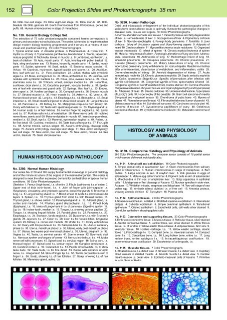
152<br />
Color Projection Slides and Photomicrographs 35 mm<br />
62. Ditto. four-cell stage 63. Ditto. eight-cell stage 64. Ditto. morula 65. Ditto.<br />
blastula 66. Ditto. gastrula 67. Giant chromosomes from Chironomus, genes and<br />
puffs 68. Human chromosomes in stage of metaphase<br />
No. 130. General Biology College Set.<br />
The selection of 75 color photomicrographs contained herein corresponds to<br />
primary and secondary school syllabuses. The series is intended to help the teacher<br />
design modern biology teaching programmes and it serves as a means of both<br />
visual and practical teaching. 75 Color Photomicrographs<br />
1. Typical animal cells 2. Amoeba proteus 3. Paramaecium 4. Hydra w.m. 5.<br />
Hydra t.s. of body 6. Trypanosoma gambiense, blood smear 7. Taenia, tapeworm,<br />
mature proglottid 8. Trichinella, larvae in muscle l.s. 9. Lumbricus, earthworm, t.s.<br />
back of clitellum 10. Apis, mouth parts 11. Apis, hind leg with pollen basket 12.<br />
Apis, sting and poison sac 13. Musca, house fly, mouth parts 14. Spider, mouth<br />
parts 15. Spider, spinneret 16. Snail, radula 17. Bacteria, mixed species 18.<br />
Volvox 19. Coprinus, mushroom, typical basidia and spores t.s. 20. Aspidium,<br />
fern, leaf with sori t.s. 21. Fern prothallium 22. Lichen, thallus with symbiotic<br />
algae t.s. 23. Moss, archegonium l.s. 24. Moss, antheridium l.s. 25. Lupinus, root<br />
nodules with symbiotic bacteria t.s. 26. Pinus, pine, ovulate cone l.s. 27. Pinus,<br />
staminate cone l.s. 28. Triticum, wheat, embryo median l.s. 29. Helianthus,<br />
sunflower, dicot stem t.s. 30. Cucurbita, pumpkin, vascular bundle t.s. 31. Epidermis<br />
of leaf with stomata and guard cells 32. Syringa, lilac, leaf t.s. 33. Elodea,<br />
stem apex l.s. 34. Hyaline cartilage t.s. 35. Compact bone t.s. 36. Smooth muscle<br />
l.s. 37. Striated muscle l.s. 38. Heart muscle l.s. 39. Artery t.s. 40. Vein t.s. 41.<br />
Human blood smear 42. Lung t.s. 43. Esophagus t.s. 44. Stomach t.s. 45. Small<br />
intestine t.s. 46. Small intestine injected to show blood vessels 47. Large intestine<br />
t.s. 48. Pancreas t.s. 49. Kidney t.s. 50. Malpighian corpuscle from kidney 51.<br />
Ovary with follicles t.s. 52. Testis with spermatogenesis t.s. 53. Thyroid gland t.s.<br />
54. Human scalp l.s. of hair follicles 55. Human finger tip sagittal l.s. 56. Spinal<br />
cord t.s. 57. Purkinje cells in t.s. of cerebellum 58. Motor nerve cells 59. Isolated<br />
nerve fibres, osmic acid 60. Motor end plates in muscle 61. Insect compound eye,<br />
median l.s. 62. Snail, eye l.s. 63. Mammal, eye median sagittal l.s. 64. Retina, t.s.<br />
for fine detail 65. Cochlea, median l.s. 66. Taste buds of tongue t.s. 67. Fish, gills<br />
t.s. 68. Animal mitosis, various stages 69. Ascaris embryology, cleavage early<br />
stage 70. Ascaris embryology, cleavage later stage 71. Sea urchin embryology,<br />
two cell stage 72. Sea urchin, four cell stage 73. Sea urchin, morula 74. Sea<br />
urchin, blastula 75. Giant chromosomes<br />
No. 3290. Human Pathology.<br />
Detail and microscopic enlargement of the individual photomicrographs of this<br />
series have been selected so as to optimally illustrate the pathological changes in<br />
diseased cells, tissues and organs. 50 Color Photomicrographs.<br />
Abnormal alterations of cells and tissues 1. Parenchymatous and fatty degeneration<br />
of liver 2. Hemosiderosis of liver 3. Glycogenosis of liver 4. Pigmentary cirrhosis<br />
of liver 5. Necrotic esophagitis 6. Foreign body granulome 7. Tonsillitis 8. Liver<br />
cirrhosis Injury of circulatory organs and blood-forming organs 9. Adiposis of<br />
heart 10. Cardiac callosity 11. Myocarditis chronica acute recidivans 12. Organized<br />
venous thrombosis 13. Infarct of spleen 14. Chronic myeloid leukemia of spleen<br />
15. Malarial melanemia of spleen Pathologic alterations of lung and liver, tuberculosis,<br />
pneumonia 16. Anthracosis of lung 17. Hemorrhagic infarct of lung 18.<br />
Influenzal pneumonia 19. Croupous pneumonia 20. Chronic pneumonia 21.<br />
Necrotic (cheesy) pneumonia 22. Miliary tuberculosis of lung 23. Chronic<br />
tuberculous pulmonary cavity with bacteria 24. Icterus hepatis Reactions or kidney<br />
after arteriosclerosis, disturbance of metabolism, and inflammation, colitis 25.<br />
Glomerularatrophy of kidney 26. Amyloid degeneration of kidney 27. Acute<br />
hemorrhagic nephritis 28. Chronic glomerulonephritis 29. Septic embolic nephritis<br />
30. Colitis dysenterica Shiga-Kruse Specific inflammations after infection with<br />
syphilis spirochaetes 31. Congenital syphilis of liver, spirochaetes silvered 32.<br />
Congenital syphilis of liver (Feuerstein liver), routine stained 33. Gumma of testicle<br />
Progressive alteration of injured tissues and organs (Hypertrophy and hyperplasia)<br />
34. Atheroma of head 35. Struma colloides 36. Undescended testicle, hyperplasia<br />
of Leydig’s cells 37. Hypertrophy of the prostate 38. Giant cell sarcoma of maxilla<br />
Benignant and malignant tumors 39. Chondroma of pubic bone 40. Myoma of<br />
uterus 41. Fibroadenoma of breast 42. Fibroepithelial mixed tumor of parotid 43.<br />
Melanosarcoma of skin 44. Spindle cell sarcoma 45. Carcinoma cervicis uteri 46.<br />
Sarcoma of testicle 47. Cystadenoma papilliferum of ovary 48. Gelatinous<br />
carcinoma of rectum 49. Lymphosarcoma mediastini 50. Metastatic carcinoma of<br />
liver.<br />
HISTOLOGY AND PHYSIOLOGY<br />
OF ANIMALS<br />
HUMAN HISTOLOGY AND PATHOLOGY<br />
No. 3280. Normal Human Histology.<br />
Our series No. 3150 and 100 supply fundamental knowledge of general histology<br />
and of the minute structure of the organs of the mammal organism. This series is<br />
designed to meet the often expressed demand for an illustration of special human<br />
conditions. 58 Color Photomicrographs.<br />
Skeleton: 1. Femur (thigh-bone), t.s. of entire 2. Fibula (calf-bone), t.s. of entire 3.<br />
Upper end of tibia (shin-bone), l.s. 4. Joint of finger with joint-capsule, l.s.<br />
Respiratory, circulatory, and lymphatic systems, endocrine glands 5. Bronchus of<br />
lung, l.s. 6. Lung showing alveoli t.s. 7. Blood smear 8. Aorta t.s. shows muscular<br />
layers 9. Spleen, t.s. 10. Thymus gland from child, t.s. with Hassall bodies 11.<br />
Thyroid gland, t.s. shows colloid 12. Parathyroid gland t.s. 13. Adrenal gland, t.s.<br />
cortex and medulla 14. Pituitary gland (Hypophysis), l.s. 15. Pineal body<br />
(Epiphysis), t.s. 16. Islets of Langerhans in t.s. of pancreas Digestive system 17.<br />
Lip, t.s. 18. Incisor tooth, median l.s. 19. Tongue, t.s. showing various papillae 20.<br />
Tongue, t.s. showing lingual follicles 21. Parotid gland t.s. 22. Pancreas t.s. 23.<br />
Esophagus, t.s. 24. Stomach, fundic region t.s. 25. Duodenum, t.s. with Brunner’s<br />
glands 26. Jejunum, t.s. 27. Colon t.s. 28. Liver, t.s. for hepatic lobes Urogenital<br />
system 29. Kidney, t.s. cortex and medulla 30. Ureter t.s. 31. Ovary with follicles<br />
t.s. 32. Ovary with Corpus luteum t.s. 33. Fallopian tube t.s. 34. Uterus, secretory<br />
phase t.s. 35. Uterus, menstrual phase t.s. 36. Uterus, early post-menstrual phase<br />
t.s. 37. Uterus, two weeks post-menstrual phase t.s. 38. Uterus, pregnant t.s. 39.<br />
Vagina t.s. 40. Testis, t.s. seminal canals 41. Sperm smear 42. Spermatic duct<br />
t.s. Nervous system and organs of sense 43. Nervus ischiadicus, t.s. 44. Motor<br />
nerve cell with processes 45. Spinal cord, t.s. cervical region 46. Spinal cord, t.s.<br />
thoracal region 47. Spinal cord, t.s. lumbar region 48. Ganglion semilunare l.s.<br />
49. Cerebral cortex t.s. 50. Cerebellum t.s. 51. Papilla circumvallata, l.s. to show<br />
taste buds 52. Taste buds, t.s. for fine detail 53. Retina with entrance of optic<br />
nerve, l.s. Integument 54. Skin of finger tip, t.s. 55. Tactile corpuscles in skin of<br />
finger l.s. 56. Scalp, showing l.s. of hair follicles 57. Scalp, showing t.s. of hair<br />
follicles 58. Mammary gland, active, t.s.<br />
No. 3150. Comparative Histology and Physiology of Animals.<br />
260 Color Photomicrographs. The complete series consists of 16 partial series<br />
which can be delivered individually also.<br />
No. 3151. Animal cell and cell division. 18 Color Photomicrographs<br />
1. Simple animal cells in salamander liver 2. Giant chromosomes from salivary<br />
gland of Chironomus 3. Human chromosomes in stage of metaphase 4. Barr<br />
bodies 5. Large oocytes in sec. of crayfish liver 6. Yolk granules in eggs of<br />
salamander 7. Mature egg cell of mammal 8. Pigment cells in skin of salamander<br />
9. Mitochondria in thin sec. of amphibian liver 10. Golgi apparatus in epithelial<br />
cells 11. Metaphase of first cleavage of Ascaris 12. Nuclear spindles in side-view,<br />
Astacus 13. Whitefish mitosis, anaphase and telophase 14. Two-cell stage of sea<br />
urchin egg 15. Amitosis (direct division) t.s. of liver cell 16. Amoeba proteus,<br />
showing amitotic division 17. Syncytium 18. Plasma cells<br />
No. 3152. Epithelial tissues. 9 Color Photomicrographs<br />
1. Squamous epithelium, isolated 2. Stratified squamous epithelium 3. Intercellular<br />
bridges 4. Cuboidal epithelium 5. Simple columnar epithelium 6. Transitional<br />
epithelium 7. Ciliated epithelium 8. Endothelial cells, cell walls silver stained 9.<br />
Glandular epithelium showing goblet cells<br />
No. 3153. Connective and supporting tissues. 20 Color Photomicrographs<br />
1. Embryonic connective tissue 2. Mucous tissue 3. Reticular tissue, silver stained<br />
4. Areolar connective tissue 5. Lattice fibres, sec. silver stained 6. White fibrous<br />
tissue, l.s. of tendon 7. Yellow elastic fibrous tissue 8. Adipose tissue, fat in situ 9.<br />
Vesicular tissue 10. Hyaline cartilage, t.s. 11. Yellow elastic cartilage, elastic<br />
fibres 12. Fibrocartilage l.s. 13. Compact bone, t.s. Haversian canals 14. Compact<br />
bone, l.s. 15. Cancellous bone, t.s. 16. Long hollow bone, entire t.s. 17. Long<br />
hollow bone, entire epiphysis l.s. 18. Intracartilaginous ossification 19.<br />
Intermembraneous ossification 20. Exoskeleton of arthropods, t.s.<br />
No. 3155. Muscular tissues. 7 Color Photomicrographs<br />
1. Striated muscle, l.s. detail view 2. Striated muscle, t.s. detail view 3. Capillary<br />
blood vessels in striated muscle 4. Smooth muscle l.s. detail view 5. Cardiac<br />
(heart) muscle l.s. detail view 6. Epithelio-muscular cells of Ascaris 7. Primitive<br />
muscle fibres of Hydra


