

BIOLOGY - microscopia.info
BIOLOGY - microscopia.info
BIOLOGY - microscopia.info

You also want an ePaper? Increase the reach of your titles
YUMPU automatically turns print PDFs into web optimized ePapers that Google loves.
Overhead Transparency Atlases<br />
125<br />
- Dicot flower bud, t.s. shows floral diagram - Arum maculatum, cuckoopint, l.s. of flower, insect trap - Lycopersicum,<br />
tomato, t.s. of flower bud shows floral diagram and axile placentation - Phaseolus, bean, t.s. of pod showing pericarp<br />
and seed - Papaver, poppy, t.s. of flower shows parietal placentation - Solanum tuberosum, potato, t.s. flower bud for<br />
floral diagram - Taraxacum, dandelion, l.s. of composite flower with tubular and ligulate florets - Taraxacum, dandelion,<br />
composite flower, color graphic design - Taraxacum, t.s. of composite flower - Cocos nucifera, coconut, endosperm t.s.<br />
- Citrus, lemon, young fruit t.s. - Triticum, wheat, t.s. of seed (grain) showing seed coat, endosperm with stored starch<br />
and embryo, entire view - Triticum, l.s. of seed (grain) showing all details, entire view, medium magnification - Triticum,<br />
l.s. through the embryo showing growing point of the stem, leaf origin, scutellum, primary root - Triticum, wheat, seed<br />
(grain), color graphic design - Zea mays, corn, grain (seed) l.s. embryo and endosperm<br />
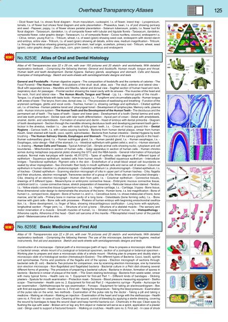
No. 8253E<br />
Atlas of Oral and Dental Histology<br />
Atlas of 40 Transparencies size 22 x 28 cm, with over 150 pictures and 20 sketch- and worksheets. With detailed<br />
explanatory textbook. - Comprising the following themes: General and foodstuffs. Human mouth, tongue and throat.<br />
Human teeth and teeth development. Dental hygiene. Salivary glands, esophagus and stomach. Cells and tissues.<br />
Examples of histopathology. Sketch and work-sheets with semidiagrammatic designs and texts<br />
General and Foodstuffs - Human digestive organs - The composition of foodstuffs and the contents of calories - The<br />
Food Pyramid –The Human Head - Articulations of the skull: skull, atlas, axis - The skull, anterior and lateral view -<br />
Skull with separated bones – Mandible and Maxilla, lateral and dorsal view - Sagittal section of human head and neck,<br />
respiratory duct. Air passages - Frontal section showing the nasal cavity with its sinuses - The muscles of the head and<br />
the neck, front and lateral view – The Human Mouth, Tongue and Throat - Lip, t.s. - Internal parts of the mouth -<br />
Tongue, t.s. of papilla foliata with taste buds - Human tongue, t.s. - Fungiform and circumvallate papilla - Human tongue<br />
with areas of taste - The larynx; front view, dorsal view, l.s. - The processes of swallowing and breathing - Function of the<br />
arytenoid cartilages, glottis and vocal cords - Trachea, human l.s. showing cartilage and epithelium - Ciliated epithelium,<br />
t.s. of trachea - Human palatine tonsil and pharyngeal tonsil - Development of lymphocytes. Memory cells, plasma<br />
cells - Human immune system - The Human Teeth and the Development of the Human Teeth - The deciduous and the<br />
permanent set of teeth - The types of teeth - Upper and lower jaws - Development of a tooth: Dental lamina and early<br />
and late tooth primordium - Dental sack with later tooth differentiation - Apical part of crown - Detail with ameloblasts,<br />
enamel, dentin, and odontoblasts - Formation of enamel and dentin - Head of embryo with dental primordia - Diagram<br />
of tooth development - Section through the mandible showing deciduous tooth and developing permanent tooth germ -<br />
Incisor in the alveolus, median l.s. - Jaw with roots of fully-grown teeth, t.s. - Crown of incisor, ground thin – Dental<br />
Hygiene - Carious tooth, l.s. with caries-causing bacteria - Bacteria from human dental plaque, smear from human<br />
mouth, Gram stained with bacilli, cocci, spirilli, spirochaetes - Bacteria from human intestine - Dental Hygiene by tooth<br />
brushing – The Human Salivary Glands, Esophagus and Stomach - The position of the salivary glands in the head -<br />
Human submaxillary gland, t.s. - Human sublingual gland, t.s. - Human parotid gland, t.s. - Human esophagus, t.s. -<br />
Esophagus, color design - Wall of the stomach, t.s. - Intestinal epithelium with goblet cellst.s. and l.s. - Human stomach,<br />
l.s. drawing – Human Cells and Tissues - Typical Animal Cell. - Simple animal cells showing nuclei, cytoplasm and cell<br />
boundaries. - Mitochondria in section of human cells. - Golgi apparatus in section of human cells - Human chromosomes<br />
during metaphase (equatorial plate) showing the GTC-and the RBA-bands - General Information of Karyotype<br />
analysis. Normal male karyotype with bands: 46,XY,GTG - Types of epithelia, color diagram of 7 different types of<br />
epithelium - Squamous epithelium, isolated cells from human mouth - Stratified squamous epithelium - Intercellular<br />
bridges - Transitional epithelium Pigment cells in the skin - Endothelium of a small blood vessel cell boundaries revealed<br />
by silver impregnation - Sex chromatin: Barr body in mouth epithelial cells and nerve cell of woman - Columnar<br />
epithelium in human intestine t.s. photomicrograph - Cuboidal epithelium t.s. photomicrograph - Ciliated epithelium, t.s.<br />
of trachea - Ciliated epithelium - Scanning electron micrograph of cilia in upper part of human trachea - Cilia, flagella<br />
and their structures, electron micrograph. Transverse section of a group of cilia; three cilia are constructed divergely -<br />
Cilia, drawing of an electron micrograph - Human skin from palm, l.s. - Columnar epithelium - Connective tissues,<br />
drawings of 6 different types - Mesenchyme or embryonic connective tissue - Embryonic mucous connective tissue,<br />
umbilical cord t.s. - Loose connective tissue, stretch preparation of mesentery. - Reticular tissue silver stained - Tendon,<br />
l.s. - Yellow elastic connective tissue (Ligamentum nuchae), t.s. - Hyaline cartilage, t.s. - Cartilage, 3 types - Bone tissue,<br />
three dimensional color design to demonstrate the structure of the bone - Human bone, t.s. low magnification - Bone of<br />
human t.s., compact bone, diagram - Bone of human t.s. and l.s. - Cancellous bone, t.s. shows trabeculae of bone, bone<br />
marrow, and fat cells - Primary bone in marrow cavity of a long bone - Osteoblasts (bone forming cells), t.s. - Bone<br />
marrow with giant cells - Bone cells with processes - Phalanx of human embryo with beginning endochondral ossification,<br />
l.s. - Bone development, l.s. finger of fetus, showing intracartilaginous ossification - Long bone with epiphysis,<br />
longitudinal section - Finger joint, l.s, - Structure of a lon g bone - Structure of a skeletal muscle - The sensory and<br />
motor innervation of a muscle - Smooth muscles of human, l.s. - Striated muscle of human, l.s. - Histopathology -<br />
Atheroma capitis, Atheroma of the head - Giant cell sarcoma of the maxilla - Ffibroepitelial mixed tumor of the parotid<br />
gland - Melanosarcoma of the skin.<br />
No. 8255E Basic Medicine and First Aid<br />
Atlas of 18 Transparencies size 22 x 28 cm, with over 76 pictures and 20 sketch- and worksheets. With detailed<br />
explanatory textbook. - Comprising the following themes: The use of the microscope, bacteria and hygiene, medical<br />
instruments, first aid and assistance. Sketch and work-sheets with semidiagrammatic designs and texts<br />
Construction of a microscope - Optical path of a microscope (path of rays) - How to prepare a microscopic slide: Blood<br />
or bacterial smear, whole mount of a zoological or botanical specimen, section of a zoological or botanical specimen -<br />
Working plan to prepare and stain a microscopic slide of a whole mount - Working plan to prepare and doubly stain a<br />
microscopic slide of a histological section (Hematoxylin-Eosine) - The different types of Bacteria. Cocci, bacilli, spirilla<br />
and spirochaetae. Forms and positions of the flagella and of the spores - Electron micrograph of sections through<br />
bacterial cells (E. coli) - Bacteria. Two pictures for comparison, one by scanning electron microscope, one by transmission<br />
electron microscope - Non-flagellated and flagellated bacteria - Bacterial culture in a Petri dish showing several<br />
different forms of growing - The procedure of preparing a bacterial culture - Bacteria in division, formation of spores in<br />
bacteria - Bacteria in smear of plaque of the teeth. – The Gram staining technology - Bacteria from waste-water, smear<br />
with many typical forms - Health care no. 1. Equipment for first-aid Part 1 - Different kinds of bandages - Sticking<br />
plasters - Spatula for mouth examination - Protection mask - Scissors - Blood pressure measuring equipment - Stethoscope<br />
- Thermometer - Health care no. 2. Equipment for first-aid Part 2 - Hypodermic syringe - Pipette - Auriscope for<br />
ear examination - Ophthalmoscope for eye examination - Forceps - Equipment for taking an electrocardiogram - Box<br />
with first-aid equipment - Health care no. 3. First aid: - Taking the temperature - Taking the blood pressure - Examination<br />
of the pulse rate on the wrist, two methods - Examination of the pulse rate by the doctor - Taking a pill and taking a<br />
medicine - Making an infusion - Making an injection - Examination of the heart and lungs with the stethoscope - Health<br />
care no. 4. First aid - In case of cuts: Cleaning of the wound, control of bleeding by applying a sterile dressing, covering<br />
the wound by bandages to keep the wound clean and keep harmful bacteria out - Chemicals in the eye: Clean eyes by<br />
flushing the eye with water - Broken arm or leg: any firm object or material will serve as a splint, application of a plaster<br />
cast - Slings used to support a fractured forearm - Walking on crutches - Health care no. 5. First aid: - In case of shock


