

BIOLOGY - microscopia.info
BIOLOGY - microscopia.info
BIOLOGY - microscopia.info

Create successful ePaper yourself
Turn your PDF publications into a flip-book with our unique Google optimized e-Paper software.
104<br />
Overhead Transparency Atlases<br />
for high and low frequencies - Detection of sound direction by the time lack between the entry of sound into the ears -<br />
Diagram of main auditory pathways. Centers of sound in the brain - Relationship of the two sets of the semicircular<br />
canals arranged in perpendicular plains - Semicircular canals, section - Ampullar crista, t.s. - Otolithic organ (macula),<br />
t.s. - Function of the vestibular system<br />
Senses of Smell, Taste, Touch, Temperature and Proprioception. - Section through nasal cavity and pharyngeal<br />
cavity - Location of the olfactory mucous membrane and air stream of the breath - Olfactory and respiratory mucous<br />
membrane of mammal t.s. - Detail view of olfactory epithelium with sensory cilia - Olfactory epithelium, electron micrograph<br />
of an ultrathin section - Nasal conchae of man and deer - Tongue of man with areas of taste - Tongue of rabbit, t.s.<br />
of papilla foliata with taste buds - Papilla foliata t.s., detail view of taste bud - Vallate papilla t.s. - Fungiform papilla t.s.<br />
- Human skin with cutaneous receptors of touch, pressure and thermal sensation - Sinus hair, l.s. and t.s. - Pacinian<br />
corpuscle - Meissner’s corpuscle from human finger - Eimer’s corpuscle from mouth of mole - Grandry’s and Herbst’s<br />
touch corpuscles from beak of duck - Sensitivity differences caused by touch-stimulation: excitation nearby or far away,<br />
weak or strong - Ruffini’s warmth receptor - Krause’s corpuscle, cold receptor - Back of human hand marked with<br />
warmth and cold reception points - Thermoreceptors of the infrared detector of rattle snake - Proprioceptors: muscle<br />
spindle and Golgi tendon apparatus. Conscious awareness of the position and movements of the joints - Muscle spindle<br />
in muscle, t.s.<br />
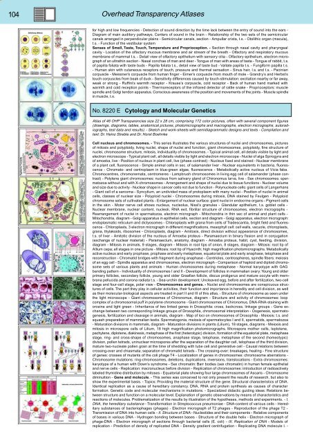
No. 8220 E Cytology and Molecular Genetics<br />
Atlas of 46 OHP Transparencies size 22 x 28 cm, comprising 172 color pictures, often with several component figures<br />
(drawings, diagrams, tables, anatomical pictures, photomicrographs and macrographs, electron micrographs, autoradiographs,<br />
test data and results). - Sketch and work-sheets with semidiagrammatic designs and texts - Compilation and<br />
text: Dr. Heinz Streble and Dr. Horst Boehnke<br />
Cell nucleus and chromosomes. - This series illustrates the various structures of nuclei and chromosomes, pictures<br />
of mitosis and polyploidy, living nuclei, shape of nuclei and function, giant chromosomes, polyploidy, fine structure of<br />
nuclei, chromosome structure, mitosis, individuality of chromosomes. - Typical animal cell, all details visible by light and<br />
electron microscope - Typical plant cell, all details visible by light and electron microscope - Nuclei of alga Spirogyra and<br />
of amoeba, live - Position of nucleus in plant cell, live (phase contrast) - Nucleus fixed and stained - Nuclear membrane<br />
of a plant cell, fluorescence - Simple animal cells in sec. of salamander liver - Nuclear equivalents in bacteria, fluorescence<br />
- Chromato- and centroplasm in blue-green algae, fluorescence - Metabolically active nucleus of Vicia faba.<br />
Chromocentres, chromonemata, centromeres - Lampbrush chromosomes in living egg cell of salamander (phase contrast)<br />
- Polytene giant chromosomes: nucleus from salivary gland of Chironomus larva, live - Sex chromosomes: spermatozoa<br />
without and with X-chromosomes - Arrangement and shape of nuclei due to tissue functions - Nuclear volume<br />
and size due to activity - Nuclear shape in cancer cells not due to function - Polynucleate cells: giant cells of Langerhans<br />
- Giant cell of a sarcoma - Syncytium, an undivided mass of protoplasm with many nuclei - Position of nuclei in animal<br />
cells, classes of nuclear size - Polyploid nuclei - Chromosomes during mitosis, DNA stained by Feulgen - Polyploid<br />
chromosome sets of cultivated plants - Enlargement of nuclear surface: giant nuclei in endocrine organs - Pigment cells<br />
in the skin - Motor nerve cell shows nucleus, nucleolus, Nissl’s granules - Glandular epithelium, t.s. goblet cells -<br />
Nuclear membrane, nuclear content, nucleoli, RNA exit, fibrillar structure of chromosomes, electron micrographs -<br />
Rearrangement of nuclei in spermatozoa, electron micrograph - Mitochondria in thin sec of animal and plant cells -<br />
Mitochondria, diagram - Golgi apparatus in epithelial cells, section and diagram - Golgi apparatus, electron micrograph:<br />
endoplasmatic reticulum and dictyosomes - Chloroplasts with grana from cells of Tradescantia, bright field and fluorescence<br />
- Chloroplasts, 3 electron micrograph in different magnifications, mesophyll cell: cell walls, vacuole, chloroplasts,<br />
grana, thylakoids, ribosomes - Chloroplasts, diagram - Amitosis, direct division without appearance of chromosomes,<br />
t.s. of liver - Amitotic division of the nucleus of Amoeba proteus - Paramaecium in binary fission and in conjugation<br />
(exchange of nuclear material) - Paramaecium, anatomy, diagram - Amoeba proteus, habit, cyst, feeding, division,<br />
diagram - Mitosis in animals, 9 stages, diagram - Mitosis in root tips of onion, 8 stages, diagram - Mitosis: root tip of<br />
Allium cepa; all stages in one picture - Mitosis: root tip of Hyacinth; high magnification photomicrographs. Metabolically<br />
active nucleus and early prophase, prophase and early metaphase, equatorial plate and early anaphase, telophase and<br />
reconstruction - Chromatid bridges with fragment during anaphase - Centrioles, centrospheres, spindle fibers: meiosis<br />
of an egg cell - Spindle apparatus and chromosomes, electron micrograph - Comparison of haploid and diploid chromosome<br />
sets of various plants and animals - Human chromosomes during metaphase - Normal karyotype with GAG<br />
banding pattern - Individuality of chromosomes I and II - Development of follicles in mammalian ovary: Young and older<br />
primary follicles, secondary follicle, young and older Graafian follicle, discus proligerus and mature oocyte with membrana<br />
pellucida and corona radiata t.s. - Sea-urchin development: Uncleaved egg, before and after fertilization, two-cell<br />
stage and four-cell stage, polar view - Chromosomes and genes. - Nuclei and chromosomes are conspicuous structures<br />
of cells. The part they play in cellular activities, their function and importance in heredity and cell division, as well<br />
as their molecular-biological aspects are treated in part II and III of this atlas. - Structure of chromosome as seen under<br />
the light microscope - Giant chromosomes of Chironomus, diagram - Structure and activity of chromosomes: loop<br />
complex of a chromosomal puff in polytene chromosome - Giant chromosomes of Chironomus, DNA-RNA-staining with<br />
orceine and light green - Inheritance of two linked genes in Drosophila: cross, backcross, linkage groups - Gene exchange<br />
between two corresponding linkage groups of Drosophila, chromosomal interpretation - Oogenesis, spermatogenesis,<br />
fertilization and cleavage in animals, diagram - Map of loci on chromosomes of Drosophila - Meiosis: t.s. and<br />
squash preparation of mammalian testis. Spermatogonia, meiosis of spermatocytes I and II, spermatids, spermatozoa<br />
- Maturation divisions in mammals, diagram - Maturation divisions in plants (Lilium), 18 stages, diagrams - Meiosis and<br />
mitosis in microspore cells of Lilium, 18 high magnification photomicrographs. Microspore mother cells, leptotene,<br />
pachytene, diplotene, diakinesis, metaphase of the first (heterotypic) division, formation of the equatorial plate, metaphase<br />
stage, ring- and cross-shape of chromosomes, anaphase stage, telophase, metaphase of the second (homeotypic)<br />
division, pollen tetrads, uninuclear microspores after the separation of the daughter cell, telophase of the third division,<br />
mature two-nucleate pollen grain at the time of shedding with tube cell and generative cell - Causal relations between<br />
crossing-over and chiasmata; separation of chromatid tetrads - The crossing-over: breakages, healing - Fine structure<br />
of genes: crosses of mutants of the coli phage T4 - Localization of genes in chromosomes: chromosome aberrations -<br />
Chromosome mutations: ring-chromosomes, deletions, duplications, inversions, translocations - Extra chromosomes:<br />
karyotype of a human with Down’s syndrome - Sex chromatin: Barr bodies (sex chromatin) in human female epithelial<br />
and nerve cells - Replication: macronucleus before division - Replication of chromosomes: introduction of radioactively<br />
labeled thymidine distribution by mitoses - Equatorial plate showing four large chromosomes of Ascaris - Chromosome<br />
diminuition - Gene and molecule. - This series was conceived to not only present the results of research, but also to<br />
show the experimental basis. - Topics: Providing the material structure of the gene. Structural characteristics of DNA.<br />
Identical replication as a cause of hereditary constancy. DNA, RNA and protein synthesis as causes of character<br />
formation. Genetic code and molecular mechanisms in mutations. - Specialized didactic guiding ideas: Relations between<br />
structure and function on a molecular level. Explanation of genetic observations by means of characteristics and<br />
reactions of molecules. Problematization of the results by illustration of the hypotheses, methods and experiments. - I.<br />
DNA, the hereditary substance - Transformation in Streptococcus pneumoniae - DNA-content of various cells - Hereditary<br />
substances of bacteriophages (phages) - Electron micrograph of T2 phages - Reproduction of the phage T2 -<br />
Transmission of DNA into human cells - II. Structure of DNA - Nucleotides and their components - Relative components<br />
of bases in various DNA - Hydrogen bonding between bases - Structure of the double helix - Electron micrograph of<br />
phage-DNA - Electron micrograph of sections through bacterial cells (E. coli) - III. Replication of DNA - Models of<br />
replication - Prediction of density of replicated DNA - Density gradient centrifugation - Replicating DNA molecule I. -


