

BIOLOGY - microscopia.info
BIOLOGY - microscopia.info
BIOLOGY - microscopia.info

Create successful ePaper yourself
Turn your PDF publications into a flip-book with our unique Google optimized e-Paper software.
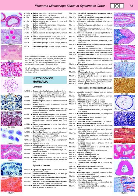
Prepared Microscope Slides in Systematic Order 61<br />
Av124d<br />
Ma101d<br />
Ma1021h<br />
Ma1033f<br />
Ma104h<br />
Ma105f<br />
Ma1055g<br />
Ma111c<br />
Ma114c<br />
Ma116d<br />
Av1245c • Gallus, cerebellum, t.s. routine stained<br />
Av1247f Gallus, cerebellum, t.s. silvered<br />
Av139d Gallus, anterior part of eye with eyelid and nictitating<br />
membrane sagittal l.s.<br />
Av140e • Gallus, posterior part of eye with retina and<br />
pecten, sagittal l.s.<br />
Av155e Gallus, chicken, horizontal sec. of the retina<br />
Av135c Gallus, cockscomb t.s.<br />
Av124d • Gallus, skin with developing feathers, horizontal<br />
l.s.<br />
Av125d • Gallus, skin with developing feathers, vertical<br />
l.s.<br />
Av126d • Gallus, unfeathered skin of foot, vertical l.s.<br />
Av211f Gallus embryology: chicken embryo, 36 hour<br />
t.s.<br />
Av212f Gallus embryology: chicken embryo, 48 hour<br />
t.s.<br />
Av213f Gallus embryology: chicken embryo, 72 hour<br />
t.s.<br />
The combination of prepared microscope slides and colour<br />
photomicrographs has decisive advantages for<br />
teaching. We have a large selection of colour photomicrographs<br />
(p. 75 – 100 in this Catalogue), for use in conjunction<br />
with our prepared microscope slides<br />
We will gladly make special offers for any slides or sets<br />
which are not listed in our catalogue. Please ask for further<br />
<strong>info</strong>rmation.<br />
HISTOLOGY OF<br />
MAMMALIA<br />
Cytology<br />
Ma101d • Simple animal cells in sec. of salamander liver<br />
showing nuclei, cell membranes and cytoplasm.<br />
For general study of the animal cell<br />
Ma102f Mitotic stages in sec. through red bone marrow<br />
of mammal<br />
Ma1023f Mitotic stages in smear of red bone marrow of<br />
mammal<br />
Ma1021h Mitotic stages in sec. of whitefish blastula<br />
showing spindles *<br />
Ma1033f • Meiotic (maturation) stages in sec. through<br />
testis of salamander, selected material showing<br />
large structures *<br />
Ma103f • Meiotic (maturation) stages in testis of mouse,<br />
sec. iron hematoxyline stained after Heidenhain<br />
Ma1031f Meiotic (maturation) stages in smear from<br />
testis of mouse, specially stained after Feulgen<br />
*<br />
Ma104h • Human chromosomes in smear from culture<br />
of blood, male *<br />
Ma1041i • Human chromosomes in smear from culture<br />
of blood, female *<br />
Ma1045f • Barr bodies (human sex chromatin) in smear<br />
from female squamous epithelium *<br />
Ma105f • Mitochondria in thin sec. of kidney or liver,<br />
specially prepared and stained<br />
Ma1055g • Golgi apparatus in sec. of spinal ganglion or<br />
other organ *<br />
Ma1058e • Pigment cells in skin<br />
Ma1061e • Storage of glycogen in liver cells, sec. stained<br />
with carmine after Best or PAS reaction<br />
Ma1063e Storage of fat in cells of costal cartilage, sec.<br />
stained with Sudan<br />
Ma1065f Secretion of fat in mammary gland, section<br />
Osmic acid stained<br />
Ma1067f • Phagocytosis in Kupffer’s star cells of the liver,<br />
sec. of mammalian liver injected with trypan<br />
blue<br />
Epithelial tissues<br />
Ma111c • Squamous epithelium, isolated cells from<br />
human mouth, smear<br />
Ma1113d Simple squamous epithelium, in sec. through<br />
the cornea from the eye<br />
Ma112c • Stratified, non-cornified squamous epithelium,<br />
in section through buccal gum<br />
Ma1121c Stratified, non-cornified squamous epithelium,<br />
in section through vagina of rabbit<br />
Ma1124d • Stratified, non-cornified squamous epithelium,<br />
in section of oesophagus<br />
Ma1125d Stratified, non-cornified squamous epithelium,<br />
t.s. pig vagina<br />
Ma1127d Stratified, cornified squamous epithelium,<br />
in vertical l.s. of human body skin<br />
Ma113d • Columnar epithelium, isolated cells from intestine<br />
w.m.<br />
Ma114c • Simple columnar epithelium, in t.s. of small<br />
intestine<br />
Ma118d<br />
Ma1142e Simple columnar epithelium, in t.s. of human<br />
gall bladder<br />
Ma1145d • Pseudostratified columnar epithelium, in<br />
sec. through epididymis<br />
Ma115d • Ciliated epithelium, isolated cells from trachea<br />
w.m.<br />
Ma116d Simple ciliated columnar epithelium, in t.s.<br />
of oviduct<br />
Ma1162d • Pseudostratified ciliated columnar epithelium,<br />
in t.s. of trachea<br />
Ma1201d<br />
Ma117e Endothelium, endothelial cells of small blood<br />
vessels in mesenterium, silver stained and w.m.<br />
Ma118d • Cuboidal epithelium, in sec. of kidney papilla<br />
Ma1182e Cuboidal epithelium, in sec. of human thyroid<br />
gland<br />
Ma120e Transitional epithelium, two section of urinary<br />
bladders showing contracted and extended<br />
Ma1202d<br />
epithelia<br />
Ma1201d • Transitional epithelium, in sec. of urinary bladder<br />
of sheep<br />
Ma1202d Goblet cells in sec. of colon, stained with mucicarmine<br />
Ma1203e Mucous glands from human intestine, colouring<br />
of goblet cells, PAS-HE<br />
Ma1204d Holocrine glands, sebaceous glands from<br />
human skin, l.s.<br />
Ma121e<br />
Ma1205c Apocrine glands, lacteal glands of sheep, sec.<br />
Ma1206e Eccrine glands, salivary gland, human, sec.<br />
Ma1207d Sweat glands in human skin, t.s.<br />
Connective and supporting tissues<br />
Ma121e • Areolar connective tissue, w.m. and stained<br />
for fibres and cells<br />
Ma122d White fibrous tissue, isolated fibres from tendon<br />
Ma123d • White fibrous tissue, l.s. of tendon<br />
Ma1231d White fibrous tissue, t.s. of tendon<br />
Ma1234f Mast cells in the Omentum majus of rat, specially<br />
stained with toluidine blue and paracarmine<br />
Ma124d Yellow elastic fibrous tissue, l.s. of Ligamentum<br />
nuchae<br />
Ma1242e • Yellow elastic fibrous tissue, t.s. of Ligamentum<br />
nuchae<br />
Ma1244d Elastic tissue, fibres teased and w.m.<br />
Ma125d Reticular tissue t.s.<br />
Ma1252f Reticular fibres, human spleen, t.s. silvered<br />
Ma126d • Embryonic connective tissue t.s.<br />
Ma127d • Mucous tissue, t.s. of navel string (umbilical<br />
cord)<br />
Ma1275f Mucous tissue, t.s. of navel string specially<br />
stained for Wharton’s jelly<br />
Ma1278d Vesicular tissue, cellular connective tissue with<br />
no intercellular substance, sec. through notochord<br />
of dogfish<br />
Ma128c • Adipose tissue, section fat removed to show<br />
the cells<br />
Ma129e • Adipose tissue, section showing fat in situ<br />
stained by sudan<br />
Ma1292e Adipose tissue, section or w.m. with fat in situ<br />
stained by osmic acid<br />
Ma1294c Brown adipose tissue of monkey, sec.<br />
Ma130c • Hyaline cartilage, t.s.<br />
Ma1302c Hyaline cartilage of cat, t.s.<br />
Ma1305d Fetal hyaline cartilage, t.s.<br />
Ma131d • Yellow elastic cartilage, section specially<br />
stained for elastic fibres<br />
Ma1312d Yellow elastic cartilage, ear of rabbit or pig,<br />
t.s.<br />
Ma132d • White fibrous cartilage, section<br />
Ma1323f Fibrous cartilage, human intervertebral disc,<br />
sec.<br />
Ma135d • Compact bone, t.s. specially prepared to show<br />
the cells and canaliculi<br />
Ma136d • Compact bone, l.s. specially prepared to show<br />
the cells and canaliculi<br />
Ma1365d • Cancellous (spongy) bone, t.s.<br />
Ma1367g Compact bone, human, non-decalcified, t.s.<br />
ground thin and mounted *<br />
Ma137e Compact bone and hyaline cartilage t.s., two<br />
sections on one slide<br />
Ma1252f<br />
Ma1278d<br />
Ma131d<br />
Ma136d<br />
Ma1365d<br />
Ma135d<br />
Ma117e<br />
Ma138e


