

BIOLOGY - microscopia.info
BIOLOGY - microscopia.info
BIOLOGY - microscopia.info

You also want an ePaper? Increase the reach of your titles
YUMPU automatically turns print PDFs into web optimized ePapers that Google loves.
Overhead Transparency Atlases<br />
121<br />
follicle mite of humans, adult specimen w.m. - Demodex folliculorum, human skin with parasites, section - Sarcoptes<br />
scabiei, penetrate through the epidermis, sec. of skin - Insecta: - Lipoptena cervi, louse fly, adult - Pediculus humanus,<br />
human louse - Phthirus pubis, pubic or crab louse - Phthirus pubis, egg attached to hair - Cimex lectularius, bed bug -<br />
Haematopinus suis, pig louse - Stomoxys, stable fly, piercing sucking mouth parts - Culex pipiens, common mosquito,<br />
pupa - Culex pipiens, posterior end of larva - Culex pipiens, adult - Culex pipiens, head and mouth parts of female and<br />
male w.m. - Culex pipiens, t.s. through the mouth parts of adult female - Culex pipiens, eggs - Anopheles, malaria<br />
mosquito, adult - Anopheles, head and mouth parts of female and male, w.m. - Pulex irritans, human flea - Xenopsylla<br />
cheopis, rat flea, carrier of the bubonic plague - Ctenocephalus canis, dog flea, adult male and female - Nosopsyllus<br />
fasciatus, rat flea - Ceratophyllus gallinulae, chicken flea - HUMAN DISEASES (PATHOLOGY): - Abnormal alterations<br />
of cells and tissues - Parenchymatous and fatty degeneration of liver - Hemosiderosis of liver - Glycogenosis of<br />
liver - Pigmentary cirrhosis of liver - Necrotic esophagitis - Foreign body granulome with hemosiderin and giant cells -<br />
Tonsillitis - Liver cirrhosis - Injury of circulatory organs and blood-forming organs - Adiposis of heart - Cardiac<br />
callosity - Myocarditis chronica acute recidivans - Organized venous thrombosis of muscle - Infarct of spleen - Chronic<br />
myeloid leukemia of spleen - Malarial melanemia of spleen - Anthracosis of lung - Pathologic alterations of lung and<br />
liver, tuberculosis, pneumonia - Cardiac callosity - Influenzal pneumonia - Croupous pneumonia - Chronic pneumonia<br />
- Necrotic (cheesy) pneumonia - Miliary tuberculosis of lung - Chronic tuberculous pulmonary cavity with bacteria -<br />
Icterus hepatis - Reaction of kidney after arteriosclerosis, disturbance of metabolism, and inflammation; colitis -<br />
Glomerular atrophy of kidney - Amyloid degeneration of kidney - Acute hemorrhagic nephritis - Chronic glomerulonephritis<br />
- Septic embolic nephritis - Colitis dysenterica Shiga-Kruse - Specific inflammations after infection with syphilis<br />
spirochaetes - Congenital syphilis of liver, spirochaetes silvered after Levaditi - Congenital syphilis of liver (Feuerstein<br />
liver), routine stained - Gumma of testicle - Progressive alteration of injured tissues and organs (Hypertrophy<br />
and hyperplasia) - Atheroma of head - Struma colloides - Undescended testicle showing hyperplasia of Leydig’s cells<br />
- Hypertrophy of prostate - Giant cell sarcoma of maxilla - Benignant and malignant tumors - Chondroma of pubic<br />
bone - Myoma of uterus - Fibroadenoma of breast - Fibroepithelial mixed tumor of parotid gland - Melanosarcoma of<br />
skin - Spindle cell sarcoma - Carcinoma cervicis uteri - Sarcoma of testicle - Cystadenoma papilliferum of ovary -<br />
Gelatinous carcinoma of rectum - Lymphosarcoma mediastini - Metastatic carcinoma of liver.<br />
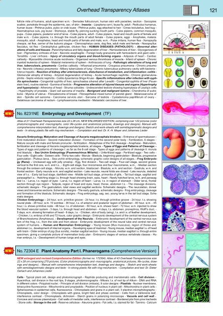
No. 8231NE Embryology and Development (TF)<br />
Atlas of 21 Overhead-Transparencies size 22 x 28 cm, NEW ENLARGED EDITION, comprising over 122 pictures (color<br />
photomicrographs and -macrographs, color life-cycles and anatomical pictures, drawings and designs). Manual with<br />
comprehensive interpretation text, drawings and designs). Sketch and work-sheets with semidiagrammatic designs and<br />
texts - In strong plastic file with ring-mechanism. – Compilation and text: Dr. K.-H. Meyer and Johannes Lieder<br />
Ascaris Embryology. Maturation and Cleavage of Ascaris megalocephala bivalens: - Entrance of spermatozoon -<br />
First maturation division - Second maturation division - Formation of the second polar body - Fertilization, 6 stages -<br />
Mature oocyte with male and female pronuclei - fertilization - Metaphase of the first cleavage - Anaphase - Maturation,<br />
fertilization and cleavage of Ascaris megalocephala bivalens, all stages. - Types of Eggs and Patterns of Cleavage: -<br />
Types of eggs and patterns of cleavage I: as far as the 8-cell stage - Types of eggs and patterns of cleavage II: morula<br />
and blastula. - Sea Urchin Embryology (Psammechinus Miliaris): - Unfertilized eggs - Fertilized eggs - Two cells -<br />
Four cells - Eight cells - Sixteen cells - Thirty-two cells - Morula - Blastula, beginning gastrulation - Blastula, progressive<br />
gastrulation - Pluteus larva, - Sea urchin embryology, schematic graphic color designs of all stages. - Frog Embryology<br />
(Rana): - Uncleaved egg with jelly envelop - Egg, first division - Two-cell stage - Four-cell stage, second groove<br />
vertical to the first one, w.m. and t.s., - Eight-cell stage, four micromeres and four macromeres, w.m., - Median section<br />
through the sixteen-cell stage, - Morula, w.m. and section, blastocoel - Blastula, w.m. and section - Gastrula, w.m. and<br />
frontal section - Early neurula w.m. and sagittal section - Late neurula, neural folds are closed - Late neurula, detailed<br />
view of t.s. - Early tail bud stage, darkfield view - Middle tail bud stage, primordia of gills, - Tail bud stage, sagittal and<br />
parasagittal l.s. - Hatching stage t.s. through head showing brain, eyes, heart - Newly hatched larva, w.m. and parasagittal<br />
l.s. - Larva, t.s. region of eyes - Larva, t.s. region of heart - Larva, t.s. in region of stomach - Older larva, frontal<br />
section through eye region - Tadpole, region of head and eyes, head, thorax, abdomen t.s. - The cleavage divisions,<br />
schematic designs - The gastrulation, total views and sagittal sections. Schematic designs - The neurulation, dorsal<br />
views and transverse sections. Schematic designs - The early gastrula, schematic designs - Frog embryology, cleavage<br />
and formation of the blastula. Schematic designs - Frog embryology, sag. sec. young larva in the tail bud stage. Schematic<br />
designs.<br />
Chicken Embryology: - 24 hour, w.m. primitive groove - 24 hour, t.s. through primitive groove - 24 hour, t.s. showing<br />
neural plate - 28 hour, w.m. 10 somites - 36 hour, t.s. of anterior and posterior region of abdomen - 40 hour, w.m. - 45<br />
hour, l.s. shows primitive node - 48 hour, t.s. of abdomen - 50 hour, w.m. shows heart - 72 hour, w.m. injected to show<br />
the blood vascular system - 3 days, t.s. through head and abdomen - 4 days, t.s. abdomen, pronephros, Wolff’s duct, -<br />
5 days, w.m. showing formation of head - 8 days, l.s. - Chicken, skin of body (wing), l.s. and t.s. of feather development,<br />
- Chicken, t.s. embryo of 48 and 72 hours, color graphic design - Embryonic development of the central nervous system<br />
of Branchiostoma (Amphioxus). - Development of the Neurula: - Embryonic development of the central nervous system<br />
of the frog, t.s., from the side and from above - Embryonic development of the neural tube and central nervous<br />
system of humans. – Human and Mammalian Embryology: - Young mouse (Mus musculus), region of thorax and<br />
abdomen t.s., development of internal organs, - Developing eyes of mammal - Young mouse, median sagittal l.s. of head<br />
with brain - Older embryo of pig (Sus scrofa), median sagittal section - Young mouse, median sagittal l.s. through entire<br />
specimen, giving a complete picture of mammalian body plan - Embryonic stages of various vertebrate classes - Human<br />
embryo, l.s. - Development of human lungs and eyes.<br />
No. 72304 E Plant Anatomy Part I. Phanerogams (Comprehensive Version)<br />
NEW enlarged and revised Comprehensive Edition (former no. 172304). Atlas of 43 Overhead-Transparencies size<br />
22 x 28 cm comprising 270 pictures. (Color photomicrographs and -macrographs, anatomical pictures, life-cycles, drawings<br />
and designs). - Manual with comprehensive interpretation text, drawings and designs. - Sketch and work-sheets<br />
with semidiagrammatic designs and texts - In strong plastic file with ring-mechanism. - Compilation and text: Dr. Dieter<br />
Gerlach and Johannes Lieder<br />
Cells - Typical plant cell, design and photomicrograph - Raphide producing and meristematic cells - Cell division -<br />
Hyacinthus, cell division in the root tips, 9 stages, photomicrographs - Mitosis: l.s. of root tip of Allium - DNA and RNA<br />
in different colors - Polyploid nuclei. - Principle of cell division (mitosis), 9 color designs - Plastids - Nuclear membrane,<br />
tetracycline fluorescence - Mitochondria and proplastids - Position of nucleus in plant cell - Mitochondria in plant cells -<br />
Spherosomes in epidermal cells, fluorescence - Chloroplasts and grana in a plant cell, 3 electron microphotographs -<br />
Chloroplasts, color design - Chloroplasts with grana, bright field and phase contrast - Cells from a Vallisneria leaf,<br />
interference contrast - Chromoplasts, dichroism - Starch grains in polarizing microscope - Vacuole and cell wall -<br />
Concave and convex plasmolysis - Cell walls of medullar cells, interference contrast - Bordered pits from pine tracheids<br />
- Stone cells - Storage in the cell - Reserve cellulose - Aleurone grains - Fat cells, t.s. stained for fat - Tannins - Calcium


