

BIOLOGY - microscopia.info
BIOLOGY - microscopia.info
BIOLOGY - microscopia.info

Create successful ePaper yourself
Turn your PDF publications into a flip-book with our unique Google optimized e-Paper software.
38<br />
Prepared Microscope Slides Sets and Series<br />
6132d<br />
6136d<br />
6151d<br />
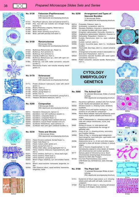
No. 6130<br />
6131c<br />
6132d<br />
6133d<br />
6134d<br />
6135d<br />
6136d<br />
No. 6150<br />
6151d<br />
6152d<br />
6153d<br />
6154d<br />
6155c<br />
6156c<br />
6157c<br />
Fabaceae (Papilionaceae)<br />
6 Microscope Slides<br />
With depictured accompanying brochure<br />
Pea (Pisum sativum), stem and petiolar tendril t.s.<br />
Pea, root with root nodules and nitrogen fixing<br />
bacteria t.s.<br />
Bean (Phaseolus vulgaris), flower bud t.s.<br />
Bean, flower bud l.s.<br />
Bean, flower showing young fruit l.s.<br />
Bean, pod with pericarp and seed t.s.<br />
Ranunculaceae<br />
Microscope Slides<br />
With depictured accompanying brochure<br />
Buttercup (Ranunculus sp.), flower l.s.<br />
Buttercup, fruit (achene) l.s.<br />
Buttercup, fruit (achene) t.s.<br />
Cowslip (Caltha sp.), fruit l.s.<br />
Buttercup (Ranunculus sp.), stem with open collateral<br />
bundles t.s.<br />
Buttercup, root with radial concentric vascular<br />
bundle t.s.<br />
Ranunculus ficaria, root nodules showing starch<br />
grains t.s.<br />
No. 6250<br />
6251d<br />
6252d<br />
6253d<br />
6254d<br />
6255d<br />
6256d<br />
6257d<br />
6258d<br />
6259d<br />
6260d<br />
6261d<br />
6262d<br />
6263d<br />
Arrangement and Types of<br />
Vascular Bundles<br />
13 Microscope Slides<br />
With depictured accompanying brochure<br />
Protostele. Psilotum, stem t.s.<br />
Actinostele. Lycopodium, stem t.s.<br />
Polystele. Pteridium, rhizome t.s. showing concentric<br />
vascular bundles with inner xylem<br />
Ectophloic siphonostele. Osmunda, rhizome t.s.<br />
Amphiphloic siphonostele. Adiantum, rhizome t.s.<br />
Dictyostele. Polypodium, rhizome t.s.<br />
Eustele. Ranunculus, stem t.s., open collateral<br />
bundles<br />
Eustele. Lamium, stem t.s.<br />
Eustele. Cucurbita pepo, stem t.s., bicollateral<br />
bundles<br />
Atactostele. Zea mays, stem t.s., closed collateral<br />
bundles<br />
Arrangement of bundles similar to atactostele in a<br />
dicot plant. Podophyllum, stem t.s.<br />
Concentric vascular bundles with outer xylem.<br />
Convallaria, rhizome t.s.<br />
Radial concentric vascular bundle. Ranunculus,<br />
root t.s.<br />
6251d<br />
6254d<br />
6255d<br />
6153d<br />
6153d<br />
No. 6170<br />
6171c<br />
6172c<br />
6173d<br />
6174d<br />
6175d<br />
6176d<br />
6177c<br />
Solanaceae<br />
7 Microscope Slides<br />
With depictured accompanying brochure<br />
Potato (Solanum tuberosum), tuber with starch<br />
grains t.s.<br />
Potato, stem t.s.<br />
Potato, young flower l.s.<br />
Potato, young flower t.s.<br />
Potato, fruit l.s.<br />
Tomato (Lycopersicum esculentum), young fruit t.s.<br />
Tobacco (Nicotiana tabacum), leaf t.s.<br />
No. 5000<br />
CYTOLOGY<br />
EMBRYOLOGY<br />
GENETICS<br />
The Animal Cell<br />
12 selected Microscope Slides of animal<br />
cytology<br />
With depictured accompanying brochure<br />
6259d<br />
6262d<br />
6202c<br />
6203d<br />
6204d<br />
6232d<br />
6236c<br />
No. 6200<br />
6201c<br />
6202c<br />
6203d<br />
6204d<br />
6205d<br />
6206d<br />
6207d<br />
6208c<br />
No. 6230<br />
6231d<br />
6232d<br />
6233b<br />
6234d<br />
6235d<br />
6236c<br />
6237c<br />
6238d<br />
6239d<br />
6240d<br />
6241d<br />
6242d<br />
Compositae<br />
8 Microscope Slides<br />
With depictured accompanying brochure<br />
Dandelion (Taraxacum), taproot t.s.<br />
Dandelion, root with lactiferous vessels l.s.<br />
Dandelion, composite flower l.s.<br />
Dandelion, composite flower t.s.<br />
Dandelion, lingulate flower isolated and w.m.<br />
Dandelion, tubular flower isolated and w.m.<br />
Sunflower (Helianthus), seed (achene) t.s.<br />
Sunflower stem with open collateral bundles t.s.<br />
Trees and Shrubs<br />
12 Microscope Slides<br />
With depictured accompanying brochure<br />
Hazel (Corylus avellana), female flower l.s.<br />
Hazel, male flower l.s.<br />
Hazel, mature pollen grains w.m.<br />
Hazel, young fruit (nut) l.s.<br />
Willow (Salix alba), young aggregate fruit l.s.<br />
Horse chestnut (Aesculus hippocastanum), petiole<br />
t.s.<br />
Horse chestnut, leaf bud t.s., shows leaf origin<br />
Horse chestnut, flower bud l.s.<br />
Horse chestnut, young fruit t.s.<br />
Beech (Fagus silvatica), sun and shadow leaves<br />
t.s.<br />
Beech, wood sections: transverse, tangential, radial<br />
Oak (Quercus robur), wood sections: transverse,<br />
tangential, radial<br />
5001c<br />
5002d<br />
5003d<br />
5004e<br />
5005d<br />
5006f<br />
5007d<br />
5008d<br />
5009e<br />
5010f<br />
5011f<br />
5012e<br />
No. 5100<br />
5101c<br />
5102d<br />
Squamous epithelium, isolated cells from human<br />
mouth. Nuclei and cytoplasm are shown<br />
Striated muscle l.s. showing nuclei, striations,<br />
myofibrils<br />
Compact bone and hyaline cartilage t.s., two<br />
sections on one slide for comparison<br />
Nerve fibres isolated, fixed and stained by osmic<br />
acid to show myeline sheaths and Ranvier’s<br />
nodes<br />
Liver of Salamandra t.s., showing simple animal<br />
cells with cellular membranes, nuclei, and<br />
cytoplasm<br />
Kidney of mouse, t.s. vital stained with<br />
trypanblue to demonstrate the storage of<br />
epithelial cells<br />
Ovary of cat, t.s. showing primary, secondary,<br />
and Graafian follicles<br />
Testis of frog, t.s. showing spermatogenesis.<br />
Spermatogonia, spermatocytes, spermatids, and<br />
mature spermatozoa<br />
Salamandra larva, t.s. of skin and other organs<br />
selected to show cell division (mitosis) in various<br />
stages<br />
Uteri of Ascaris megalocephala, t.s. iron<br />
hematoxyline stained to show details of meiosis<br />
with chromosomes and nuclear spindles<br />
Salivary gland of Chironomus larva. Giant<br />
chromosomes showing large chromomeres.<br />
Stained for DNA after Feulgen<br />
Ova from Psammechinus (sea urchin).<br />
Unfertilized ova, fertilized ova, early cleavage<br />
stages<br />
The Plant Cell<br />
12 selected Microscope Slides of plant<br />
cytology<br />
With depictured accompanying brochure<br />
Epidermis of Allium cepa (onion), w.m. showing<br />
simple plant cells with cell walls, nuclei and<br />
cytoplasm<br />
Root tips of Allium cepa l.s. showing cell division<br />
(mitosis) in all stages, clearly stained<br />
5005d<br />
5007d<br />
5010f<br />
5011f<br />
5101c<br />
6242d<br />
5102d


