

BIOLOGY - microscopia.info
BIOLOGY - microscopia.info
BIOLOGY - microscopia.info

Create successful ePaper yourself
Turn your PDF publications into a flip-book with our unique Google optimized e-Paper software.
102<br />
Overhead Transparency Atlases<br />
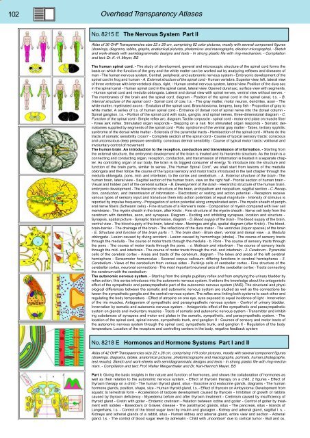
No. 8215 E The Nervous System Part II<br />
Atlas of 36 OHP Transparencies size 22 x 28 cm, comprising 82 color pictures, mostly with several component figures<br />
(drawings, diagrams, tables, graphs, anatomical pictures, photomicro- and macrographs, electron micrographs). - Sketch<br />
and work-sheets with semidiagrammatic designs and texts - In strong plastic file with ring-mechanism. - Compilation<br />
and text: Dr. K.-H. Meyer, BS<br />
The human spinal cord. - The study of development, general and microscopic structure of the spinal cord forms the<br />
basis on which the function of the grey and the white matter can be worked out by analyzing reflexes and diseases of<br />
man - The human nervous system. Central, peripheral, and autonomic nervous system - Embryonic development of the<br />
spinal cord in frog and human - A. External structure of the spinal cord - Human vertebra. Superior view, left, lateral view<br />
of three vertebrae with intervertebral discs, right. - Human central nervous system, lateral view. Position of the dura sac<br />
in the spinal canal - Human spinal cord in the spinal canal, lateral view. Opened dural sac, surface view with segments.<br />
- Human spinal cord and medulla oblongata. Lateral and dorsal view with spinal nerves, ventral view without nerves. -<br />
The membranes of the brain and the spinal cord, diagram - Position of the spinal cord in the spinal canal, t.s. - B.<br />
Internal structure of the spinal cord - Spinal cord of cow, t.s. - The gray matter, motor neuron, dendrites, axon - The<br />
white matter, myelinated axons - Evolution of the spinal cord. Branchiostoma, lamprey, bony fish - Proportion of gray to<br />
white matter. A series of t.s. of human spinal cord - Entrance of dorsal root of spinal nerve into the dorsal column -<br />
Spinal ganglion, l.s. - Portion of the spinal cord with roots, ganglia, and spinal nerves, three-dimensional diagram - C.<br />
Function of the spinal cord - Simple reflex arc, diagram. Tactile corpuscle - spinal cord - motor end plate on muscle fiber<br />
- Knee jerk reflex. Stimulated organ responds - Stepping on a nail. Not stimulated organ responds - Somatic dermatomes<br />
supplied by segments of the spinal cord - Polio: syndrome of the ventral gray matter - Tabes, tertiary syphilis:<br />
syndrome of the dorsal white matter - Sclerosis of the pyramidal tracts - Hemisection of the spinal cord - Where do the<br />
tracts of somatic sensibility cross? - Complete section of the spinal cord - Course of typical sensory tracts: conscious<br />
and unconscious deep pressure sensibility, conscious dermal sensibility - Course of typical motor tracts: volitional and<br />
involuntary control of movement<br />
The human brain. An introduction to the reception, conduction and transmission of <strong>info</strong>rmation. - Starting from<br />
the external structure, the embryonic development of the brain is treated and its hierarchic structure. As the brain is a<br />
connecting and conducting organ, reception, conduction, and transmission of <strong>info</strong>rmation is treated in a separate chapter.<br />
As controlling organ of our body, the brain is its biggest consumer of energy. To introduce into the structure and<br />
function of the brain parts, similar to series „The Human Spinal Cord“, we shall start from lesions of the medulla<br />
oblongata and then follow the course of the typical sensory and motor tracts introduced in the last chapter through the<br />
medulla oblongata, pons, mid- and interbrain, to the cortex and cerebellum. - A. External structure of the brain - The<br />
human brain, lateral view - Sagittal section of the human brain, view on the right half - Frontal section of human brain -<br />
Visual and hidden part of the cerebral surface - B. Development of the brain - Hierarchic structure of the human brain,<br />
embryonic development - The hierarchic structure of the brain, archipallium and neopallium, sagittal section - C. Reception,<br />
conduction, and transmission of <strong>info</strong>rmation - Electrotonic or resting and action potential - Receptors receive<br />
various types of sensory input and transduce them into action potentials of equal magnitude - Intensity of stimulus is<br />
reported by impulse frequency - Propagation of action potential along unmyelinated axon - The myelin sheath of peripheral<br />
nerve fibers (Schwann cells) - Fine structure of a Ranvier’s node - Composition of myelin compared with liver cell<br />
membrane - The myelin sheath in the brain, after Krstic - Fine structure of the myelin sheath - Nerve cell body from the<br />
cerebrum with dendrites, axon, and synapses. Diagram - Exciting and inhibiting synapses, location and structure -<br />
Synapsis, spatial picture - Synaptic transmission, diagram - D. Blood supply of the brain - The blood supply of the brain,<br />
ventral view - The blood supply of the brain, lateral view - Meninges and glia, spatial diagram (after Krstic) - The bloodbrain-barrier<br />
- The drainage of the brain - The reflections of the dura mater - The ventricles (liquor spaces) of the brain<br />
- E. Structure and function of the brain parts - 1. The brain stem - Brain stem, ventral and dorsal view - a. Medulla<br />
oblongata - Lesion caused by diving accident - Lesion caused by hemorrhage (stroke) - The course of sensory tracts<br />
through the medulla - The course of motor tracts through the medulla - b. Pons - The course of sensory tracts through<br />
the pons - The course of motor tracts through the pons. - c. Midbrain and interbrain - The course of sensory tracts<br />
through the mid- and interbrain - The course of motor tracts through the mid- and interbrain - 2. Cerebrum - Pyramidal<br />
cells of the cerebral cortex - Areas and tracts of the cerebrum, diagram - The lobes and areas of the left cerebral<br />
hemisphere - Sensomotor homunculus - Severed corpus callosum: differing functions in cerebral hemispheres - 3.<br />
Cerebellum - Views of the cerebellum from various sides - Purkinje cells of cerebellar cortex - Fine structure of the<br />
cerebellar cortex, neuronal connections - The most important neuronal arcs of the cerebellar cortex - Tracts connecting<br />
the cerebrum with the cerebellum<br />
The autonomic nervous system. - Starting from the simple pupillary reflex and from emptying the urinary bladder by<br />
reflex action, this series introduces into the autonomic nervous system. It widens the knowledge about the antagonistic<br />
effect of the sympathetic and parasympathetic part of the autonomic nervous system (ANS). The structural and physiological<br />
differences between the somatic and autonomic nervous system are studied as well as the connections between<br />
the sympathetic ganglia and the central nervous system. The reflex arcs linking both systems to each other and<br />
regulating the body temperature. - Effect of atropine on one eye, eyes exposed to equal incidence of light - Innervation<br />
of the iris muscles. Antagonism of sympathetic and parasympathetic nervous system - Control of urinary bladder.<br />
Innervation by somatic and autonomic nervous system. - Antagonistic effect of the sympathetic and parasympathetic<br />
system on glands and involuntary muscles - Tracts of somatic and autonomic nervous system - Transmitter and inhibiting<br />
substances of synapses and motor end plates in the somatic, sympathetic, and parasympathetic system. - The<br />
location of the spinal cord, spinal nerves, sympathetic trunk, and ganglion II - Courses of sensory and motor tracts of<br />
the autonomic nervous system through the spinal cord, sympathetic trunk, and ganglion II - Regulation of the body<br />
temperature. Location of the receptors and controlling centers in the body, negative feedback system<br />
No. 8218 E Hormones and Hormone Systems Part I and II<br />
Atlas of 42 OHP Transparencies size 22 x 28 cm, comprising 116 color pictures, mostly with several component figures<br />
(drawings, diagrams, tables, anatomical pictures, photomicrographs and macrographs, portraits, human photographs,<br />
test results). Sketch and work-sheets with semidiagrammatic designs and texts - In strong plastic file with ring-mechanism.<br />
- Compilation and text: Prof. Walter Mergenthaler and Dr. Karl-Heinrich Meyer, BS<br />
Part I: Giving the basic insights in the nature and function of hormones, and shows the collaboration of hormones as<br />
well as their relation to the autonomic nervous system. - Effect of thyroxin therapy on a child, 2 figures - Effect of<br />
thyroxin therapy on a child - The human thyroid gland, situs - Exocrine and endocrine glands, diagrams - The human<br />
hormone glands, position, shape, size - Human thyroid gland, t.s. - Effect of thyroxin on Ambystoma: Development from<br />
aquatic to terrestrial form - Acceleration of tadpole development caused by thyroxin - Inhibition of growth of rabbits<br />
caused by thyroxin deficiency - Myxedema before and after thyroxin treatment - Cretinism caused by insufficiency of<br />
thyroid gland - Cretin with goiter - Endemic cretinism - Relation between iodine and goiter - Control of goiter by treatment<br />
with iodides - Basedow’s or Graves’ disease - The parathyroid glands, situs - The pancreas, situs - Islands of<br />
Langerhans, t.s. - Control of the blood sugar level by insulin and glucagon - Kidney and adrenal gland, sagittal l. s. -<br />
Kidneys and adrenal glands of a rabbit, situs - Human kidney and adrenal gland, entire view and section - Adrenal<br />
gland, t.s. - The control of blood sugar level by adrenalin - Child with „moonface“ due to cortical tumor - Bull and ox,


