

BIOLOGY - microscopia.info
BIOLOGY - microscopia.info
BIOLOGY - microscopia.info

Create successful ePaper yourself
Turn your PDF publications into a flip-book with our unique Google optimized e-Paper software.
118<br />
Overhead Transparency Atlases<br />
medulla oblongata. Lateral and dorsal view with spinal nerves, ventral view without nerves - Brain stem with cranial<br />
nerves, ventral and dorsal view - Human vertebrae. Superior view and lateral view of three vertebrae with intervertebral<br />
discs - Human central nervous system, lateral view. Position of the dura sac in the spinal canal - Spinal cord, t.s. routine<br />
stained - Spinal cord, t.s. silver stained - Gray matter of spinal cord, t.s. showing nerve cell bodies - White matter of<br />
spinal cord, t.s. showing nerve fibers - Portion of the spinal cord with roots, ganglia, and branches of spinal nerves,<br />
three-dimensional diagram - Sympathetic ganglion, human t.s. - Pseudounipolar neuron (T-cell) from spinal ganglion -<br />
Peripheral nerve, human sciatic nerve, t.s., low magnification - Peripheral nerve, bundle from sciatic nerve, t.s., medium<br />
magnification - Peripheral nerve, nerve fibers, t.s., high magnification, axons and medullary sheaths - Peripheral nerve,<br />
teased material of osmic acid fixed material with Ranvier’s nodes - Optic nerve, human t.s. - Motor nerve cells, smear<br />
from spinal cord of ox shows nerve cells and their appendages - Various shapes of human neurons, 5 figures - Nerve<br />
cell body from the cerebrum with dendrites, axon, and synapses. Diagram - Synapsis, spatial picture - Motor nerve<br />
endings, muscle stained with gold chloride showing the motor end plates - Motor end plates (neuromuscular junction),<br />
diagram, 2 figures - The human eye. Eyeball, eye muscles, eyelid, sagittal section - Eye, anterior part with iris, ciliary<br />
body, cornea, l.s. - Eye, posterior part with retina and entrance of optic nerve, l.s. - Retina from eye, t.s. for all details -<br />
Human retina, chief synaptic connections, color diagrammatic design - Cornea from eye, human t.s. - Eyelid of cat, t.s.<br />
showing Meibomian gland - Cochlea (internal ear) from guinea pig, l.s. showing organ of Corti - Morphology of the<br />
human ear. Ear concha, external auditory canal, middle ear, internal ear - Organ of Corti, two color spatial diagrams -<br />
Olfactory region from nose of rabbit, t.s. - Olfactory epithelium with sensory cilia, t.s. detail view - Tongue of rabbit with<br />
papilla foliata shows abundant taste buds, t.s. - Papilla foliata t.s., detail view of taste bud, high magnification - Vallate<br />
papilla of the human tongue t.s., detail view - Tactile hairs with blood sinus, l.s. - Tactile hairs with blood sinus, t.s. -<br />
Touch corpuscles in human skin, t.s. - Grandry corpuscles in t.s. through beak of duck - Pacinian corpuscles in mesentery<br />
or pancreas - Meissner’s corpuscle from human finger - Krause’s corpuscle, cold receptor<br />
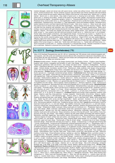
No. 8237 E Zoology (Invertebrates) (TB)<br />
Atlas of 26 Overhead-Transparencies size 22 x 28 cm, comprising over 165 pictures (color photomicrographs and -<br />
macrographs, color life-cycles and anatomical pictures).Manual with comprehensive depictured interpretation text. In<br />
strong plastic file with ring-mechanism. - Sketch and work-sheets with semidiagrammatic designs and texts - Compilation<br />
and text: Dr. K.-H. Meyer and Johannes Lieder<br />
Protozoa Amoeba proteus - Amoeba, color design showing habit, cyst, feeding, division, - Euglena, green flagellate -<br />
Euglena, habit, division, conjugation and formation of cysts. Color design - Radiolaria, mixed - Foraminifera, mixed -<br />
Trypanosoma gambiense, habit and division, color design and blood smear - Ceratium, dinoflagellates - Noctiluca<br />
miliaris - Plasmodium falciparum, tertian malaria, blood smear - Plasmodium berghei, smear with asexual and sexual<br />
stages - Eimeria stiedae, coccidiosis, section of liver - Paramecium, ciliate, anatomy, color design, living specimen,<br />
macro- and micronuclei stained, binary fission, conjugation and structure of the surface - Vorticella, a stalked ciliate,<br />
living specimen - Porifera Sponge of the sycon type, schematic design and t.s. - Spongilla, fresh water sponge, t.s. -<br />
Sycon, spicules w.m. - Euspongia, commercial sponge, skeleton - Coelenterata Hydra, fresh water polyp, anatomy and<br />
reproduction, color design - Hydra with bud, transverse section, nematocysts, color design - Hydra, l.s. of a specimen<br />
with ingested food - Polyp and medusa (Obelia), life cycle and development - Obelia hydroid, vegetative and reproductive<br />
polyps - Obelia medusa, jellyfish, w.m. - Aurelia, ephyra w.m. - Actinia, sea anemone, t.s. and l.s. - Platyhelminthes<br />
Fasciola hepatica (Distomum hepaticum) liver fluke, digestive, reproductive, excretory, nervous systems, color designs<br />
- Fasciola hepatica, t.s. body - Planaria, w.m. general body plan of a flatworm and t.s. - Taenia saginata, tapeworm, t.s.<br />
of proglottids and color design - Taenia saginata, mature proglottid w.m. - Moniezia, tapeworm, scolex - Echinococcus<br />
granulosus, dog tapeworm, adult specimen with scolex and proglottids and t.s. of cyst with scolices - Nemathelminthes<br />
Ascaris, roundworm, t.s., color design - Ascaris, t.s. of adult female - Nemathelminthes, body plan of male and female,<br />
color design - Trichinella spiralis, section and flat mount of infected muscle with encysted larvae - Enterobius vermicularis<br />
(Oxyuris), pin worm - Nereis, t.s. of body - Hirudo medicinalis, medicinal leech, t.s. - Lumbricus, earthworm,<br />
copulating specimens, reproduction and t.s. color design - Lumbricus, t.s. of body back of the clitellum - Lumbricus,<br />
anterior end 1.-9., 9.-16. and 16.-23. segment l.s. - Crustacea Daphnia and Cyclops, small crustaceans, anatomy, color<br />
design - Daphnia, water flea, living specimen - Cyclops, copepods - Artemia salina, brine shrimp - Astacus, crayfish,<br />
gills t.s. - Astacus, ovary t.s. with developing eggs - Astacus, testis t.s. with spermatogenesis - Arachnida Spider,<br />
anatomy and general body plan, color design - Spider, sagittal l.s. and t.s with book lung - Spider, entire young specimen<br />
- Spider, leg with comb, survey and high magnification - Spider, spinneret - Scorpion, w.m. for all details and<br />
sagittal section - Scorpion, poison gland - Varroa, parasitic mite of bees - Argas persicus, tick - Ixodes, tick, six legged<br />
larva - Dermanyssus gallinae, chicken mite - Tyroglyphus farinae, mite from meal - Insecta Musca domestica, house fly,<br />
head and mouth parts w.m., t.s. and color design - Apis mellifica, honey bee, mouth parts w.m., t.s. and color design -<br />
House fly, sucking tube, scanning electron micrograph - Blatta, cockroach, anatomy and general body plan of insects,<br />
color design - Periplaneta or Blatta, cockroach, biting mouth parts of a herbivore w.m. - Periplaneta, head and mouth<br />
parts, color design - Blatta, cockroach, adult female, dorsal view, reproductive organs, color design - Pieris, butterfly,<br />
clubbed antenna - Bombyx mori, silk moth, feathered antenna - Melolontha, cockchafer, laminate antenna - Pieris,<br />
butterfly, mouth parts with sucking tube - Musca domestica, structure of the leg with pulvilli - Apis mellifica, honey bee,<br />
structure of the leg, color design - Apis mellifica, posterior leg with pollen basket and foreleg with antenna cleaner -<br />
Melolontha, cockchafer, digging leg - Apis mellifica, anterior and posterior wings - Musca domestica, wing with halters<br />
- Spiracle (stigma) of insect, surface view and section, color design and w.m. - Pieris, butterfly, wing showing arrangement<br />
of scales, w.m. and design - Trachea from insect, showing elastic spiral threads, w.m. and design - Periplaneta,<br />
cockroach, upper and lower wings - Tracheal gills, of May fly nymph, design - Compound eye of an insect, histology,<br />
schematic design - Head with brain and eyes of an insect t.s., design - Compound eye, t.s. through head of honey bee<br />
- Cornea, showing facets - Compound eye of Melolontha, showing superposed insect eye, l.s. - Grasshopper, testis t.s.<br />
with spermatogenesis - Sting of honey bee, anatomy and function, color design and w.m. - Apis mellifica, honey bee, t.s.<br />
of abdomen of drone showing testis - Apis mellifica, t.s. of abdomen of queen showing ovaries - Anopheles, malaria<br />
mosquito, adult female - Anopheles, head and mouth parts of female and male - Culex pipiens, common mosquito, adult<br />
female - Culex pipiens, head and mouth parts of female and female - Drosophila, fruit fly, adult - Flea, anatomy, color<br />
design - Ctenocephalus canis, dog flea, adult male - Pulex irritans, human flea, adult female and male - Pediculus<br />
humanus, human louse - Cimex lectularius, bed bug - Aphidae sp., plant lice - Mollusca Chiton, a primitive mollusk, t.s.<br />
through the body - Alloteuthis, cuttlefish, entire young specimen - Octopus, cuttlefish, section through sucking tube -<br />
Cuttlefish, anatomy and general body plan, color graphic design - Camera eye of cuttlefish (Sepia), l.s. - Mya arenaria,<br />
t.s. and l.s. of gills - Anodonta, mussel, complete t.s. - Mussel (clam), anatomy and general body plan, color design -<br />
Snail, anatomy and general body plan, color design - Snail, typical t.s. - Helix pomatia, Roman snail, hermaphrodite<br />
gland (ovotestis) t.s. - Echinodermata Echinus, sea urchin, reproduction, color design - Asterias, horizontal and sagittal<br />
l.s. with internal organs, color design - Asterias, arm t.s. and color design - Sea urchin embryology, uncleaved<br />
fertilized egg, 2-cell stage, 4-cell stage, 8-cell stage, morula, blastula, gastrula and pluteus larva - Acrania, Fish,<br />
Amphibians, Reptiles and Birds Branchiostoma lanceolatum, block diagram combined with t.s. and l.s., color design<br />
- Branchiostoma, typical t.s. shows gills, liver and gonads - Scyllium, dogfish, region of head and gills, t.s. - Fresh water<br />
fish, abdominal region t.s. - Cyprinus, carp, blood smear - Fish scales, cycloid, ctenoid and placoid scales - Salamandra<br />
larva, head with eyes t.s. - Rana, frog, blood smear - Rana, stomach t.s. gastric glands - Rana, small intestine (duodenum)<br />
t.s. - Rana, lung t.s., simple baglike lung - Rana, kidney t.s., Malpighian corpuscles - Rana, testis showing spermatogenesis<br />
t.s. - Rana, ovary with developing eggs t.s. - Rana, skin with skin glands, l.s. - Lacerta, lizard, skin with<br />
scales, l.s. - Lacerta, lizard, lung t.s. - Gallus, chicken, blood smear - Gallus, lung t.s. showing bird lung with parabronchi<br />
- Bird feathers, construction and function, color design - Bird feathers, wing and down


