

BIOLOGY - microscopia.info
BIOLOGY - microscopia.info
BIOLOGY - microscopia.info

You also want an ePaper? Increase the reach of your titles
YUMPU automatically turns print PDFs into web optimized ePapers that Google loves.
Overhead Transparency Atlases<br />
101<br />
placenta, diagram - Fetus in uterus showing placenta, umbilical cord, and amniotic cavity - Full term baby in maternal<br />
abdomen, normal cephalic presentation - Beginning of birth, entrance of amniotic sac into the birth canal<br />
Germ development of man and animals. - Starting with the fertilization of the egg and the fusion of the two haploid<br />
nuclei, the various types of egg and corresponding types of cleavage are shown. The gastrulation, neurulation and<br />
formation of germ layers in Branchiostoma, frog and human beings are then illustrated. - Fertilization of the Ascaris egg,<br />
entering of a sperm I. The beginning of embryonic development - fertilization - Fertilization of Ascaris egg, entrance of<br />
spermatozoon in the oocyte - Mature oocyte of Ascaris with male and female pronuclei, each nucleus contains two<br />
chromosomes. - II. Cleavage - Metaphase of the first cleavage of Ascaris, equatorial plate in side view shows chromosomes,<br />
spindle fibers, centrioles - Telophase of the first cleavage of Ascaris, division of the cell body - Total equal<br />
cleavage: 2-, 4-, 8-cell stage, morula - Types of eggs and patterns of cleavage I: as far as the 8-cell stage - Types of<br />
eggs and patterns of cleavage II: morula and blastula - Blastula of sea urchin (Echinus), after total equal cleavage -<br />
Blastula of frog (Rana), after total unequal cleavage - Insect, blastula after superficial cleavage - III. Gastrulation -<br />
Gastrulation of sea urchin, Echinus, diagram - Gastrula of sea urchin, Echinus, photomicrograph - IV. Neurulation -<br />
Organogenesis in frog and chicken - Neurulation in Amphioxus, t.s. diagram - Neurulation in frog, antero-lateral and<br />
dorsal view, diagram - Neurulation in frog, t.s. - Neurula of frog, t.s. - Neurula of frog, mid-dorsal region, t.s., detail -<br />
Neurula of chicken, t.s. - Chicken embryo 33 hours of incubation, l.s. - Frog embryo, tail bud stage, l.s. - Frog embryo,<br />
tail bud stage, t.s. - Frog larva, 3 days after hatching, l.s. - Frog larva after hatching, t.s. - Frog larva, t.s. of heart region<br />
- Chicken embryo, 48-hours, t.s. - Chicken embryo, 72-hours, l.s. - Chicken embryo, 72-hours chick, embryonic disc with<br />
circular system injected - Chicken, older embryo, l.s. - V. Organogenesis in humans, Summary - Median l.s. through a<br />
human embryo - Development of the human heart, t.s. of three stages - External changes in the human heart, ventral<br />
view - Development of human lungs, t.s. of 6 weeks old embryo - Stages of human pulmonary development - Development<br />
of the human eyes, four stages - Head of mammalian embryo, sagittal section showing eyes - Mammalian embryo,<br />
median sagittal section of whole body with primordia of organs<br />
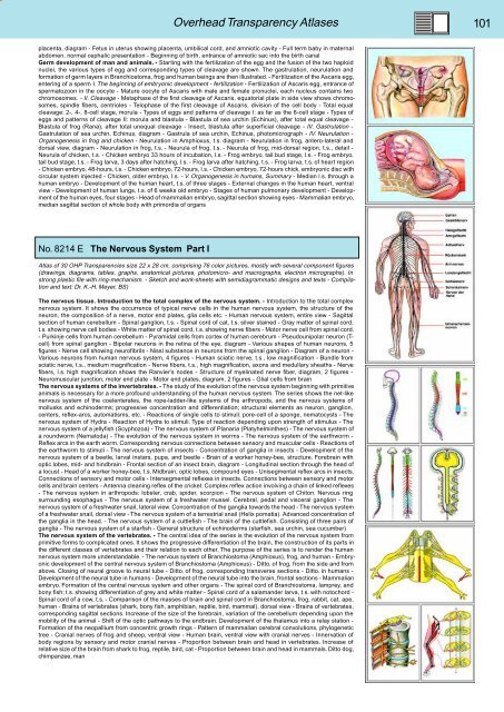
No. 8214 E The Nervous System Part I<br />
Atlas of 30 OHP Transparencies size 22 x 28 cm, comprising 76 color pictures, mostly with several component figures<br />
(drawings, diagrams, tables, graphs, anatomical pictures, photomicro- and macrographs, electron micrographs). In<br />
strong plastic file with ring-mechanism. - Sketch and work-sheets with semidiagrammatic designs and texts - Compilation<br />
and text: Dr. K.-H. Meyer, BS)<br />
The nervous tissue. Introduction to the total complex of the nervous system. - Introduction to the total complex<br />
nervous system. It shows the occurrence of typical nerve cells in the human nervous system, the structure of the<br />
neuron, the composition of a nerve, motor end plates, glia cells etc. - Human nervous system, entire view - Sagittal<br />
section of human cerebellum - Spinal ganglion, t.s. - Spinal cord of cat, t.s. silver stained - Gray matter of spinal cord,<br />
t.s. showing nerve cell bodies - White matter of spinal cord, t.s. showing nerve fibers - Motor nerve cell from spinal cord.<br />
- Purkinje cells from human cerebellum - Pyramidal cells from cortex of human cerebrum - Pseudounipolar neuron (Tcell)<br />
from spinal ganglion - Bipolar neurons in the retina of the eye, diagram - Various shapes of human neurons, 5<br />
figures - Nerve cell showing neurofibrils - Nissl substance in neurons from the spinal ganglion - Diagram of a neuron -<br />
Various neurons from human nervous system, 4 figures - Human sciatic nerve, t.s., low magnification - Bundle from<br />
sciatic nerve, t.s., medium magnification - Nerve fibers, t.s., high magnification, axons and medullary sheaths - Nerve<br />
fibers, l.s. high magnification shows the Ranvier’s nodes - Structure of myelinated nerve fiber, diagram, 2 figures -<br />
Neuromuscular junction, motor end plate - Motor end plates, diagram, 2 figures - Glial cells from brain<br />
The nervous systems of the invertebrates. - The study of the evolution of the nervous system beginning with primitive<br />
animals is necessary for a more profound understanding of the human nervous system. The series shows the net-like<br />
nervous system of the coelenterates, the rope-ladder-like systems of the arthropods, and the nervous systems of<br />
mollusks and echinoderms; progressive concentration and differentiation; structural elements as neuron, ganglion,<br />
centers, reflex-arcs, automatisms, etc. - Reactions of single cells to stimuli: pore-cell of a sponge, nematocysts - The<br />
nervous system of Hydra - Reaction of Hydra to stimuli. Type of reaction depending upon strength of stimulus - The<br />
nervous system of a jellyfish (Scyphozoa) - The nervous system of Planaria (Platyhelminthes) - The nervous system of<br />
a roundworm (Nematoda) - The evolution of the nervous system in worms - The nervous system of the earthworm -<br />
Reflex arcs in the earth worm. Corresponding nervous connections between sensory and muscular cells - Reactions of<br />
the earthworm to stimuli - The nervous system of insects - Concentration of ganglia in insects - Development of the<br />
nervous system of a beetle, larval instars, pupa, and beetle - Brain of a worker honey-bee, structure. Forebrain with<br />
optic lobes, mid- and hindbrain - Frontal section of an insect brain, diagram - Longitudinal section through the head of<br />
a locust - Head of a worker honey-bee, t.s. Midbrain, optic lobes, compound eyes - Unisegmental reflex arcs in insects.<br />
Connections of sensory and motor cells - Intersegmental reflexes in insects. Connections between sensory and motor<br />
cells and brain centers - Antenna cleaning reflex of the cricket. Complex reflex action involving a chain of linked reflexes<br />
- The nervous system in arthropods: lobster, crab, spider, scorpion - The nervous system of Chiton. Nervous ring<br />
surrounding esophagus - The nervous system of a freshwater mussel. Cerebral, pedal and visceral ganglion - The<br />
nervous system of a freshwater snail, lateral view. Concentration of the ganglia towards the head - The nervous system<br />
of a freshwater snail, dorsal view - The nervous system of a terrestrial snail (Helix pomatia). Advanced concentration of<br />
the ganglia in the head. - The nervous system of a cuttlefish - The brain of the cuttlefish. Consisting of three pairs of<br />
ganglia - The nervous system of a starfish - General structure of echinoderms (starfish, sea urchin, sea cucumber)<br />
The nervous system of the vertebrates. - The central idea of the series is the evolution of the nervous system from<br />
primitive forms to complicated ones. It shows the progressive differentiation of the brain, the construction of its parts in<br />
the different classes of vertebrates and their relation to each other. The purpose of the series is to render the human<br />
nervous system more understandable. - The nervous system of Branchiostoma (Amphioxus), frog, and human - Embryonic<br />
development of the central nervous system of Branchiostoma (Amphioxus) - Ditto. of frog, from the side and from<br />
above. Closing of neural groove to neural tube - Ditto. of frog, corresponding transverse sections - Ditto. in humans -<br />
Development of the neural tube in humans - Development of the neural tube into the brain, frontal sections - Mammalian<br />
embryo. Formation of the central nervous system and other organs - The spinal cord of Branchiostoma, lamprey, and<br />
bony fish; t.s. showing differentiation of grey and white matter - Spinal cord of a salamander larva, t.s. with notochord -<br />
Spinal cord of a cow, t.s. - Comparison of the masses of brain and spinal cord in Branchiostoma, frog, rabbit, cat, ape,<br />
human - Brains of vertebrates (shark, bony fish, amphibian, reptile, bird, mammal), dorsal view - Brains of vertebrates,<br />
corresponding sagittal sections. Increase of the size of the forebrain, variation of the cerebellum depending upon the<br />
mobility of the animal - Shift of the optic pathways to the endbrain. Development of the thalamus into a relay station -<br />
Formation of the neopallium from concentric growth rings - Pattern of mammalian cerebral convolutions, phylogenetic<br />
tree - Cranial nerves of frog and sheep, ventral view - Human brain, ventral view with cranial nerves - Innervation of<br />
body regions by sensory and motor cranial nerves - Proportion between brain and head in vertebrates. Increase of<br />
relative size of the brain from shark to frog, reptile, bird, cat - Proportion between brain and head in mammals. Ditto dog,<br />
chimpanzee, man


