

BIOLOGY - microscopia.info
BIOLOGY - microscopia.info
BIOLOGY - microscopia.info

Create successful ePaper yourself
Turn your PDF publications into a flip-book with our unique Google optimized e-Paper software.
Overhead Transparency Atlases<br />
97<br />
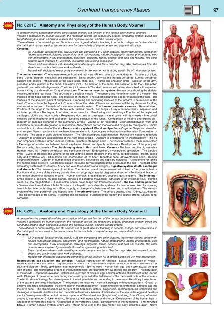
No. 8201E Anatomy and Physiology of the Human Body. Volume I<br />
A comprehensive presentation of the construction, biology and function of the human body in three volumes.<br />
Volume I comprises the human skeleton, the muscular system, the respiratory organs, circulatory system, blood and<br />
lymphatic organs, heart and blood vessels, the digestive system, and the urinary organs.<br />
These atlases of human biology and life science are of great value for teaching in schools, colleges and universities, in<br />
the training of nurses, medical technicians and for the students of physiotherapy and physical education.<br />
Contents:<br />
• 36 Overhead-Transparencies, size 22 x 28 cm, comprising 110 color pictures, mostly with several component<br />
figures (anatomical pictures, photomicro- and macrographs, nature photographs, human photographs, electron<br />
micrographs, X-ray photographs, drawings, diagrams, tables, scenes, test data and results). The color<br />
pictures were prepared by university illustrators specializing in this field.<br />
• Sketch and work-sheets with semidiagrammatic designs and texts. Teacher may take photocopies from the<br />
sheets and use for classroom work and tests.<br />
• Manual with depictured explanatory comments for the teacher. All in strong plastic file with ring-mechanism.<br />
The human skeleton - The human skeleton, front and rear view - Fine structure of bone, diagram - Structure of a long<br />
bone - Joints: diagram, hinge, ball-and-socket joint - Spinal column, cervical and thoracic vertebrae - Lumbar vertebrae,<br />
sacrum and coccyx - Articulations of the skull: skull, atlas, axis - Thorax and shoulder girdle - Skeleton of the arm,<br />
pronation and supination of the hand - The elbow joint - The skeleton of the hand - The skeleton of the foot - The pelvic<br />
girdle with and without its ligaments - The knee joint, menisci - The skull, anterior and lateral view - Skull with separated<br />
bones - X-ray of a dislocation - X-ray of a fracture - The human muscular system - Human body showing the skeletal<br />
muscles, front and rear views - The structure of a skeletal muscle - The sensory and motor innervation of a muscle - The<br />
muscles of the head and the neck - The muscles of the trunk - The superficial and the deeper muscles of the back - The<br />
muscles of the shoulder, pairs of antagonists - Pronating and supinating muscles of the forearm - The muscles of the<br />
hand - The muscles of the leg and foot - The muscles of the pelvis - Flexors and extensors of the leg - Muscles for lifting<br />
and lowering the arm - Example of a complex muscular action - The human respiratory system - General view -<br />
Position of the lungs in the thorax. Thorax with trachea, bronchi, and lungs - X-ray of human thorax, inspirated and<br />
expirated position - The larynx; front view, dorsal view, l.s. - Swallowing and breathing - Function of the arytenoid<br />
cartilages, glottis and vocal cords - Respiratory duct and air passages - Nasal cavity with its sinuses - Intercostal<br />
muscles during inspiration and expiration - Detailed structure of the lungs - Comparison of inspired and expired air -<br />
Diagram of gaseous exchange in the pulmonary alveoli - Volume of air respirated - Connection between work and<br />
respiration per minute - Regulation of respiration- Absorption of carbon monoxide and oxygen by hemoglobin - Smoke<br />
and sulphur dioxide-content of the air - The circulatory system I: Blood and lymphatic Organs - Shape and size of an<br />
erythrocyte - Serum reactions to show hereditary relationship - Leucocytes with phagocyted bacteria - Composition of<br />
the blood - The steps of blood clotting, diagram - The AB0 blood group determination - Positive and negative reactions<br />
- Diagram to understand agglutination of the AB0-blood groups - Diagram to understand Rh-incompatibility - The human<br />
lymphatic system - Human immune system - Structure of a lymph node - The vascular system of the human spleen<br />
- Exchange of substances between blood capillaries, tissue, and lymph capillaries - Development of lymphocytes.<br />
Memory cells, plasma cells - The circulatory system II: Heart and blood Vessels - The heart and the big vessels -<br />
Human heart, l.s. - Arterio-ventricular and semilunar valves - Endocardium, myocardium, epicardium - The cardiac<br />
cycle - Cycle of pressure and volume of the left ventricle. Blood pressure in the aorta, cardiac sounds - Heart, pulmonary<br />
and systemic loop - Stimulation and coordination of the heart. Sinoatrial node, atrioventricular node - Human<br />
electrocardiogram - Diagram of human blood circulation. Big vessels and capillary networks - Arrangement for taking<br />
the human blood pressure - Diagram to explain the pulse during reduction of the pressure in the bag - The heart in the<br />
circulatory system of vertebrates - Artery and vein, three-dimensional designs - Digestive system. Mouth, esophagus<br />
and stomach - The human organs of nutrition - The deciduous and the permanent set of teeth - The types of teeth -<br />
Position and structure of the salivary glands - Human esophagus, spatial diagram and section - Position and fixation of<br />
the human abdominal digestive organs. - Human stomach, spatial diagram, sections, gastric glands - The intestine -<br />
Small intestine, sections, mucous glands, principle of peristaltic movement - Structure of an intestinal villus - Human<br />
colon, l.s., low magnification - Human colon, spatial color design and transverse section - The liver and the pancreas<br />
- General structure of a liver lobule- Structure of a hepatic cord - Vascular systems of a liver lobule - Liver, t.s. showing<br />
liver lobules, bile ducts, diagram - Blood supply, exchange of substances of liver and small intestine - The venous<br />
system of the liver, portal vein and hepatic vein - The urinary organs - The urinary organs, situs - Kidney, l.s., diagram<br />
- The blood vessels of the kidney - Nephron and glomerulus - Function of the kidney, the course of renal tubules, renal<br />
corpuscle.<br />
No. 8202E Anatomy and Physiology of the Human Body. Volume II<br />
A comprehensive presentation of the construction, biology and function of the human body in three volumes.<br />
Volume I comprises the human skeleton, the muscular system, the respiratory organs, circulatory system, blood and<br />
lymphatic organs, heart and blood vessels, the digestive system, and the urinary organs.<br />
These atlases of human biology and life science are of great value for teaching in schools, colleges and universities, in<br />
the training of nurses, medical technicians and for the students of physiotherapy and physical education.<br />
Contents:<br />
• 32 Overhead-Transparencies, size 22 x 28 cm, comprising 101 color pictures, mostly with several component<br />
figures (anatomical pictures, photomicro- and macrographs, nature photographs, human photographs, electron<br />
micrographs, X-ray photographs, drawings, diagrams, tables, scenes, test data and results). The color<br />
pictures were prepared by university illustrators specializing in this field.<br />
• Sketch and work-sheets with semidiagrammatic designs and texts. Teacher may take photocopies from the<br />
sheets and use for classroom work and tests.<br />
• Manual with depictured explanatory comments for the teacher. All in strong plastic file with ring-mechanism.<br />
Reproduction, sex education and genetics - Asexual reproduction of Amoeba - Sexual reproduction of Hydra -<br />
Reproduction of the sea urchin - Reproduction in fishes - The reproductive organs of the human male; lateral view of<br />
situs and diagram - Testis, epididymis, spermatogenesis - Spermatozoa - Human hair, egg, and spermatozoa; comparison<br />
of sizes - The reproductive organs of the human female; lateral and front view of situs and diagram - The maturation<br />
of the oocyte - Oogenesis, ovulation, fertilization, cleavage of fertilized egg, and implantation of blastocyst in the uterine<br />
wall - Changes of the endometrium during menstrual cycle and after fertilization - The menstrual cycle of the woman -<br />
The fertilization of the egg, first development in the fallopian tube and imbedding in the uterus - Hereditary transmission<br />
of the sex and sex-linked inheritance - The human chromosomes - Normal karyotype with banding pattern - Growth of<br />
embryo and fetus in the uterus - Full term baby in maternal abdomen - Beginning of birth, entrance of amniotic sac into<br />
the birth canal - The chromosomes as carriers of the hereditary factors - Oogenesis, spermatogenesis fertilization and<br />
cleavages in animals - Fertilization and maturation divisions in Ascaris - Fertilization of the sea urchin egg and development<br />
- Development of the central nervous systems of Branchiostoma (Amphioxus) and frog, from. Closing of neural<br />
groove to neural tube - Chicken embryo, 48 hour, t.s. with neural tube and chorda - Development of the human heart -<br />
Graduation of vertebrate hearts - Graduation of the vertebrate lungs - Development of the human eye - The nervous<br />
tissue - Human nervous system, entire view - Motor nerve cells of the gray matter, cell body, dendrites, axon - Nerve


