BIOLOGY - microscopia.info
BIOLOGY - microscopia.info
BIOLOGY - microscopia.info
You also want an ePaper? Increase the reach of your titles
YUMPU automatically turns print PDFs into web optimized ePapers that Google loves.
106<br />
Overhead Transparency Atlases<br />
fibers isolated, showing myeline sheaths and Ranvier’s nodes - Simple animal cells in liver, t.s. with cellular membranes,<br />
nuclei, and cytoplasm - Electron micrograph of a liver cell showing nucleus, mitochondria, cytosomes, lysosomes,<br />
dictyosomes, glycogen - Phagocytosis in Kupffer’s star cells of the liver, t.s. - Ovary of cat, t.s. showing primary, secondary,<br />
and Graafian follicles - Fallopian tube with embedded egg (oocyte), t.s. high magnification detail - Testis of frog, t.s.<br />
showing spermatogenesis. Spermatogonia, spermatocytes, spermatids, and mature spermatozoa - Testis of crayfish,<br />
t.s. showing meiosis and spermatogenesis - Animal mitosis, color graphic design with 9 different stages - Reduction<br />
division during spermatogenesis in human and animals, all stages, color graphic design - Giant chromosomes in the<br />
salivary gland of Chironomus larva, with large chromomeres. Stained for DNA - Giant chromosomes of Chironomus,<br />
color graphic design - Human chromosomes in smear from culture of blood - Karytype of human chromosomes -<br />
Lampbrush chromosomes of diplotene stage in living egg cell of salamander (phase contrast) - Uteri of Ascaris megalocephala,<br />
t.s. to show details of meiosis with chromosomes and nuclear spindles - Barr bodies (sex chromatin) in<br />
female squamous epithelium - Pigment cells in skin - Storage of glycogen in liver cells, sec. - Nucleus of an amoeba, live<br />
microphotograph - Mitochondria in thin sec. of kidney or liver, specially prepared and stained - Mitochondria, fine<br />
structure, color schematic design - Golgi apparatus in sec. of spinal ganglion - Golgi apparatus, fine structure, color<br />
schematic design - Ova from Psammechinus (sea urchin). Fertilized ovum - Ova from Psammechinus (sea urchin). Twocell<br />
stage - Ova from Psammechinus (sea urchin). Four-cell stage - Ova from Psammechinus (sea urchin). Eight-cell<br />
stage - Inheritance of two linked genes in Drosophila: cross, backcross, linkage groups - Gene exchange between two<br />
corresponding linkage groups of Drosophila, chromosomal interpretation - Drosophila genetics, adult wild type, w.m. -<br />
Drosophila, “barr eye” mutant, w.m. - Drosophila, “brown eye” mutant, w.m. - Drosophila, “vestigial wing” mutant, w.m. -<br />
Drosophila, “white eye” mutant, w.m.<br />
Plant Cells and Genetics: - Typical Plant Cell, showing all details visible by light and electron microscope in different<br />
colors - Electron micrograph of a plant cell with nucleus, cell walls, vacuoles, mitochondria, endoplasmatic reticulum,<br />
plasmodesma and chloroplasts - Epidermis of Allium (onion), w.m. showing simple plant cells with cell walls, nuclei and<br />
cytoplasm - Stem apex and meristematic tissue of Elodea, l.s. showing growing zone and leaf origin - Wood of Tilia<br />
macerated and w.m. showing wood cells, vessels and fibers - Root tips of Allium l.s. showing cell division (mitosis) in all<br />
stages: - Mitosis: root tip; interphase (resting stage) - Mitosis: root tip; early prophase - Mitosis: root tip; late prophase -<br />
Mitosis: root tip; early metaphase - Mitosis: root tip; equatorial plate of metaphase - Mitosis: root tip; early anaphase -<br />
Mitosis: root tip; telophase - Mitosis: root tip; reconstruction - Maturation divisions (meiosis and mitosis) in the pollen<br />
mother cells of Lilium, 18 stages, color design - Pollen mother cells of Lilium. Early prophase (leptotene) first division<br />
(meiosis) showing chromosomes as fine threads - Pollen mother cells of Lilium. Later prophase (diakinesis) of first<br />
division (meiosis) Shortening of chromosomes - Pollen mother cells. Metaphase and anaphase of first division (meiosis)<br />
showing nuclear spindles and contracted chromosomes - Pollen mother cells. Second division, interkinesis, four cells<br />
stage - Plasmodesmata, in t.s. of palm seed - Mitochondria, thin l.s. of Allium root tips stained to show the mitochondria<br />
- Fruit of Pyrus (pear) t.s. showing stone cells (scelerenchyma) - Tuber of Solanum (potato) t.s. shows cork and starch<br />
grains - Cucurbita (pumpkin) l.s. of stem showing vascular bundles with sieve tubes, spiral and annular vessels, sclerenchyma<br />
fibers - Ricinus endosperm t.s. showing aleurone grains - Ovary of Lilium (lily), t.s. showing arrangement of<br />
ovules and embryosac - Spirogyra, green alga, showing conjugation stages and formation of zygotes.<br />
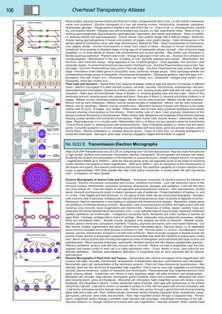
No. 8222 E Transmission Electron Micrographs<br />
Atlas of 24 OHP Transparencies size 22 x 28 cm, comprising over 120 individual pictures. They are made from extremely<br />
high quality, faultless and instructive transmission electron micrographs. All micrographs are marked with letters<br />
facilitating the location and interpretation of the important or special structures. Greatly enlarged electron micrographs<br />
- magnification 50000 up to 100000 x - show the ultra-structures of the cell organelles as far as the range of macromolecules.<br />
Electron micrographs of lower magnification - 5000 up to 30000 x - give an impression of the microstructure of<br />
the tissues and organs, their specific performance and functions. The resolution capability of a modern electron microscope<br />
is approximately 1000 times greater than that of the optical microscope. In strong plastic file with ring-mechanism.<br />
- Compilation: Dr. Heinz Streble<br />
Electron Micrographs of Animal Cells and Tissues. - Techniques: production of ultra-thin sections for electron microscopy<br />
- Electron microscope: composition and function, refraction and lenses - Liver cell: distinctive marks of fine<br />
structure; nucleus, mitochondria, cytosomes, lysosomes, dictyosomes, glycogen, gall capillaries - Liver cell: fine structure<br />
of an animal cell - Liver cell: details of cell organelles and endoplasmatic reticulum - Skin: desmosomes, tonofilaments,<br />
microvilli and fissures for lymph in stratum spinosum cells of epidermis - Ciliated epithelium of trachea: t.s. and<br />
l.s. of cilia - Cilia, flagella and their structures: t.s. of a group of cilia; three cilia are constructed divergently - Secretory<br />
cells: exocrine cells of pancreas, endoplasmatic reticulum and dictyosomes as origin-structures of digestion enzymes -<br />
Ribosomes: fixed on membranes or free floating in cytoplasm the ribosomes form designs - Resorption: simple columnar<br />
epithelium of intestine showing microvilli - Resorption: cells of proximal tubule of kidney; the highly active cells with<br />
numerous long microvilli, basal invaginations and mitochondria - Glomerulus of kidney, details: capillary loops and<br />
podocytes; the barrier between blood and primary urine - Lung: epithelial layer of pneumocytes, basement membrane<br />
capillary epithelium and erythrocytes - Collagenous connective tissue: fibroblasts and matrix bundles of banded collagen<br />
fibrils - Cartilage: cartilage cells in matrix of cartilage - Bone, osteocytes: long cytoplasmatic processes, collagen<br />
fibrils and mineralized matrix - Smooth muscle: elongated units showing two kinds of filaments - Skeletal muscle,<br />
striated: plasma membrane, sarcoplasm, myofibrils, T-tubules, segments and bands, actin and myosin filaments - Cardiac<br />
muscle, striated: segmentation and bands, mitochondria, intercalated discs - Nervous tissue: t.s. of myelinated<br />
axons and non-myelated axons within grooves of Schwann’s cells - Nervous tissue: l.s. of axon, neurofilaments, microtubules,<br />
vesicles, mitochondria, Schwann’s cell with node of Ranvier - Neuro-muscular synapses in skeletal muscle: the<br />
junction shows vesicles in presynaptic component and junctional folds that reach the myofibrils in postsynaptic component<br />
- Blood: mature erythrocytes including homogeneous mass of hemoglobin, and erythroblast with large nucleus and<br />
polyribosomes - Blood: granular leukocytes, eosinophils: lobulated nucleus and disc-shaped cytoplasmatic granules -<br />
Olfactory epithelium: sensory cells with cilia, mucous cells w. microvilli - Retina: rod cells in longitudinal view; the outer<br />
segment and banded rootlet of each cell is a highly specialized cilium - Ovary: details of ovum, zona pellucida and<br />
follicular epithelium. - Testicles; spermatogenic epithelium: in longitudinal view an early spermatid and an matured<br />
spermatozoon<br />
Electron Micrographs of Plant Cells and Tissues. - Typical plant cells: electron micrograph of low magnification with<br />
nucleus, cell walls, vacuoles, mitochondria, dictyosomes, endoplasmatic reticulum, plasmodesma and chloroplasts -<br />
Meristematic plant cell: representation of the membrane systems - Plant cell: three dimensional reconstruction - Meristematic<br />
plant cell: fine structures of organelles; high magnified - Cell of root tip: very high magnified cut-out showing<br />
cell wall, plasma membrane, clusters of ribosomes and microtubules - Plasmodesmata: high magnified electron micrograph<br />
showing details - Cytokinesis and mitosis in early telophase stage: cell plate formation and phragmoplast -<br />
Mesophyll cell: cell walls, large vacuole, chloroplasts, grana of plastids, starch and nucleotides - Mesophyll cell: chloroplast<br />
showing starch, grana and thylakoids - Mesophyll cell: chloroplast; highly magnified cut-out with details in grana,<br />
thylakoids, and ribosomes in stroma - Cuticle: epidermal cuticle of petiole, cutin layer with residual wax on the surface<br />
and primary cell wall - Leaf stoma: section cut parallel to surface of a leaf, with two guard cells and two subsidiary cells<br />
- Leaf stoma: transverse sections through stoma cells - Gland cells: section through a gland from leaf of privet showing<br />
gland cells and a stalk cell - Root: central cylinder, transverse section showing Casparian strips, endodermis, cortex,<br />
gas spaces, pericycle, sieve tubes and tracheids - Root: high magnified section through a Casparian strip - Primary<br />
xylem: longitudinal section through a primitive xylem element with secondary, ring-shaped thickenings of the wall -<br />
Vascular cambium: t.s. through cambium of a woody stem; low magnification - Vascular cambium, detail: cambial initial


