

BIOLOGY - microscopia.info
BIOLOGY - microscopia.info
BIOLOGY - microscopia.info

You also want an ePaper? Increase the reach of your titles
YUMPU automatically turns print PDFs into web optimized ePapers that Google loves.
Overhead Transparency Atlases<br />
119<br />
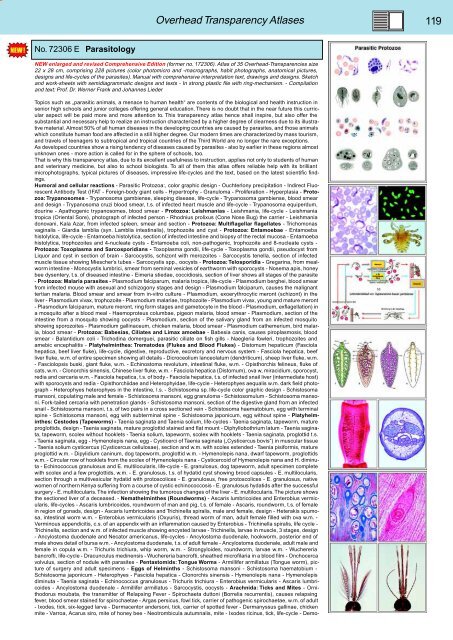
No. 72306 E Parasitology<br />
NEW enlarged and revised Comprehensive Edition (former no. 172306). Atlas of 35 Overhead-Transparencies size<br />
22 x 28 cm, comprising 228 pictures (color photomicro and -macrographs, habit photographs, anatomical pictures,<br />
designs and life-cycles of the parasites). Manual with comprehensive interpretation text, drawings and designs. Sketch<br />
and work-sheets with semidiagrammatic designs and texts - In strong plastic file with ring-mechanism. - Compilation<br />
and text: Prof. Dr. Werner Frank and Johannes Lieder<br />
Topics such as „parasitic animals, a menace to human health“ are contents of the biological and health instruction in<br />
senior high schools and junior colleges offering general education. There is no doubt that in the near future this curricular<br />
aspect will be paid more and more attention to. This transparency atlas hence shall inspire, but also offer the<br />
substantial and necessary help to realize an instruction characterized by a higher degree of clearness due to its illustrative<br />
material. Almost 50% of all human diseases in the developing countries are caused by parasites, and those animals<br />
which constitute human food are affected in a still higher degree. Our modern times are characterized by mass tourism,<br />
and travels of teenagers to subtropical and tropical countries of the Third World are no longer the rare exceptions.<br />
As developed countries show a rising tendency of diseases caused by parasites - also by earlier in these regions almost<br />
unknown ones - more action is called for in the sphere of schools, too.<br />
That is why this transparency atlas, due to its excellent usefulness to instruction, applies not only to students of human<br />
and veterinary medicine, but also to school biologists. To all of them this atlas offers reliable help with its brilliant<br />
microphotographs, typical pictures of diseases, impressive life-cycles and the text, based on the latest scientific findings.<br />
Humoral and cellular reactions - Parasitic Protozoa:, color graphic design - Ouchterlony precipitation - Indirect Fluorescent<br />
Antibody Test (IFAT - Foreign-body giant cells - Hypertrophy - Granuloma - Proliferation - Hyperplasia - Protozoa:<br />
Trypanosomes - Trypanosoma gambiense, sleeping disease, life-cycle - Trypanosoma gambiense, blood smear<br />
and design - Trypanosoma cruzi blood smear, t.s. of infected heart muscle and life-cycle - Trypanosoma equiperdum,<br />
dourine - Apathogenic trypanosomes, blood smear - Protozoa: Leishmanias - Leishmania, life-cycle - Leishmania<br />
tropica (Oriental Sore), photograph of infected person - Rhodnius prolixus (Cone Nose Bug) the carrier - Leishmania<br />
donovani, Kala Azar, from infected spleen, smear and section - Protozoa: Multiflagellar flagellates - Trichomonas<br />
vaginalis - Giardia lamblia (syn. Lamblia intestinalis), trophozoite and cyst - Protozoa: Entamoebae - Entamoeba<br />
histolytica, life-cycle - Entamoeba histolytica, section of infected intestine and biopsy of the rectal mucosa - Entamoeba<br />
histolytica, trophozoites and 4-nucleate cysts - Entamoeba coli, non-pathogenic, trophozoite and 8-nucleate cysts -<br />
Protozoa: Toxoplasma and Sarcosporidians - Toxoplasma gondii, life-cycle - Toxoplasma gondii, pseudocyst from<br />
Liquor and cyst in section of brain - Sarcocystis, schizont with merozoites - Sarcocystis tenella, section of infected<br />
muscle tissue showing Miescher’s tubes - Sarcocystis spp., oocysts - Protozoa: Telosporidia - Gregarina, from mealworm<br />
intestine - Monocystis lumbrici, smear from seminal vesicles of earthworm with sporocysts - Nosema apis, honey<br />
bee dysentery, t.s. of diseased intestine - Eimeria stiedae, coccidiosis, section of liver shows all stages of the parasite<br />
- Protozoa: Malaria parasites - Plasmodium falciparum, malaria tropica, life-cycle - Plasmodium berghei, blood smear<br />
from infected mouse with asexual and schizogony stages and design - Plasmodium falciparum, causes the malignant<br />
tertian malaria. Blood smear and smear from in-vitro culture - Plasmodium, exoerythrocytic meront (schizont) in the<br />
liver - Plasmodium vivax, trophozoite - Plasmodium malariae, trophozoite - Plasmodium vivax, young and mature meront<br />
- Plasmodium falciparum, mature meront, ring form stages and gametocyte in the blood - Plasmodium, exflagellation) in<br />
a mosquito after a blood meal - Haemoproteus columbae, pigeon malaria, blood smear - Plasmodium, section of the<br />
intestine from a mosquito showing oocysts - Plasmodium, section of the salivary gland from an infected mosquito<br />
showing sporozoites - Plasmodium gallinaceum, chicken malaria, blood smear - Plasmodium cathemerium, bird malaria,<br />
blood smear - Protozoa: Babesias, Ciliates and Limax amoebae - Babesia canis, causes piroplasmosis, blood<br />
smear - Balantidium coli - Trichodina domerguei, parasitic ciliate on fish gills - Naegleria fowleri, trophozoites and<br />
amebic encephalitis - Platyhelminthes: Trematodes (Flukes and Blood Flukes) - Distomum hepaticum (Fasciola<br />
hepatica, beef liver fluke), life-cycle, digestive, reproductive, excretory and nervous system - Fasciola hepatica, beef<br />
liver fluke, w.m. of entire specimen showing all details - Dicroceolium lanceolatum (dendriticum), sheep liver fluke, w.m.<br />
- Fasciolopsis buski, giant fluke, w.m. - Echinostoma revolutum, intestinal fluke, w.m. - Opisthorchis felineus, fluke of<br />
cats, w.m. - Clonorchis sinensis, Chinese liver fluke, w.m. - Fasciola hepatica (Distomum), ova w, miracidium, sporocyst,<br />
redia and cercaria w.m. - Fasciola hepatica, t.s. of body - Fasciola hepatica, t.s. of infected snail liver (intermediate host)<br />
with sporocysts and redia - Opisthorchiidae and Heterophyidae, life-cycle - Heterophyes aequalis w.m. dark field photograph<br />
- Heterophyes heterophyes in the intestine, l.s. - Schistosoma sp. life-cycle color graphic design - Schistosoma<br />
mansoni, copulating male and female - Schistosoma mansoni, egg granuloma - Schistosomulum - Schistosoma mansoni.<br />
Fork-tailed cercaria with penetration glands - Schistosoma mansoni, section of the digestive gland from an infected<br />
snail - Schistosoma mansoni, t.s. of two pairs in a cross sectioned vein - Schistosoma haematobium, egg with terminal<br />
spine - Schistosoma mansoni, egg with subterminal spine - Schistosoma japonicum, egg without spine - Platyhelminthes:<br />
Cestodes (Tapeworms) - Taenia saginata and Taenia solium, life-cycles - Taenia saginata, tapeworm, mature<br />
proglottids, design - Taenia saginata, mature proglottid stained and flat mount - Diphyllobothrium latum - Taenia saginata,<br />
tapeworm, scolex without hooklets - Taenia solium, tapeworm, scolex with hooklets - Taenia saginata, proglottid t.s.<br />
- Taenia saginata, egg - Hymenolepis nana, egg - Cysticerci of Taenia saginata („Cysticercus bovis“) in muscular tissue<br />
- Taenia solium cysticercus (Cysticercus cellulosae), section and w.m. with scolex extended - Taenia pisiformis, mature<br />
proglottid w.m. - Dipylidium caninum, dog tapeworm, proglottid w.m. - Hymenolepis nana, dwarf tapeworm, proglottids<br />
w.m. - Circular row of hooklets from the scolex of Hymenolepis nana - Cysticercoid of Hymenolepis nana and H. diminuta<br />
- Echinococcus granulosus and E. multilocularis, life-cycle - E. granulosus, dog tapeworm, adult specimen complete<br />
with scolex and a few proglottids, w.m. - E. granulosus, t.s. of hydatid cyst showing brood capsules - E. multilocularis,<br />
section through a multivesicular hydatid with protoscolices - E. granulosus, free protoscolices - E. granulosus, native<br />
women of northern Kenya suffering from a course of cystic echinococcosis - E. granulosus hydatids after the successful<br />
surgery - E. multilocularis. The infection showing the tumorous changes of the liver - E. multilocularis. The picture shows<br />
the sectioned liver of a deceased. - Nemathelminthes (Roundworms) - Ascaris lumbricoides and Enterobius vermicularis,<br />
life-cycles - Ascaris lumbricoides, roundworm of man and pig, t.s. of female - Ascaris, roundworm, t.s. of female<br />
in region of gonads, design - Ascaris lumbricoides and Trichinella spiralis, male and female, design - Heterakis spumosa,<br />
intestinal worm w.m. - Enterobius vermicularis (Oxyuris), thread worm of man, adult female filled with ova w.m. -<br />
Verminous appendicitis, c.s. of an appendix with an inflammation caused by Enterobius - Trichinella spiralis, life cycle -<br />
Trichinella, section and w.m. of infected muscle showing encysted larvae - Trichinella, larvae in muscle, 3 stages, design<br />
- Ancylostoma duodenale and Necator americanus, life-cycles - Ancylostoma duodenale, hookworm, posterior end of<br />
male shows detail of bursa w.m. - Ancylostoma duodenale, t.s. of adult female - Ancylostoma duodenale, adult male and<br />
female in copula w.m. - Trichuris trichiura, whip worm, w.m. - Strongyloides, roundworm, larvae w.m. - Wuchereria<br />
bancrofti, life-cycle - Dracunculus medinensis - Wuchereria bancrofti, sheathed microfilaria in a blood film - Onchocerca<br />
volvulus, section of nodule with parasites - Pentastomids: Tongue Worms - Armillifer armillatus (Tongue worm), picture<br />
of surgery and adult specimens - Eggs of Helminths - Schistosoma mansoni - Schistosoma haematobium -<br />
Schistosoma japonicum - Heterophyes - Fasciola hepatica - Clonorchis sinensis - Hymenolepis nana - Hymenolepis<br />
diminuta - Taenia saginata - Echinococcus granulosus - Trichuris trichiura - Enterobius vermicularis - Ascaris lumbricoides<br />
- Ancylostoma duodenale - Armillifer armillatus - Sarcocystis, oocysts - Arachnida: Ticks and Mites - Ornithodorus<br />
moubata, the transmitter of Relapsing Fever - Spirochaeta duttoni (Borrelia recurrentis), causes relapsing<br />
fever, blood smear stained for spirochaetae - Argas persicus, fowl tick, carrier of pathogenic spirochaetae, w.m. of adult<br />
- Ixodes, tick, six-legged larva - Dermacentor andersoni, tick, carrier of spotted fever - Dermanyssus gallinae, chicken<br />
mite - Varroa, Acarus siro, mite of honey bee - Neotrombicula autumnalis, mite - Ixodes ricinus, tick, life-cycle - Demo-


