

BIOLOGY - microscopia.info
BIOLOGY - microscopia.info
BIOLOGY - microscopia.info

Create successful ePaper yourself
Turn your PDF publications into a flip-book with our unique Google optimized e-Paper software.
40<br />
Prepared Microscope Slides Sets and Series<br />
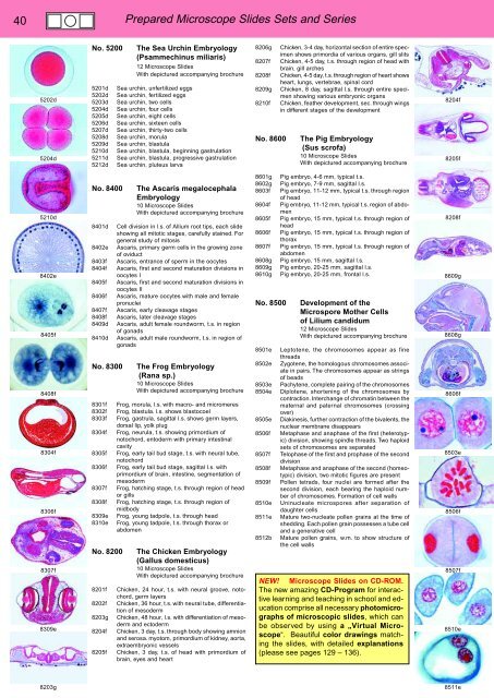
5202d<br />
5204d<br />
No. 5200<br />
5201d<br />
5202d<br />
5203d<br />
5204d<br />
5205d<br />
5206d<br />
5207d<br />
5208d<br />
5209d<br />
5210d<br />
5211d<br />
5212d<br />
The Sea Urchin Embryology<br />
(Psammechinus miliaris)<br />
12 Microscope Slides<br />
With depictured accompanying brochure<br />
Sea urchin, unfertilized eggs<br />
Sea urchin, fertilized eggs<br />
Sea urchin, two cells<br />
Sea urchin, four cells<br />
Sea urchin, eight cells<br />
Sea urchin, sixteen cells<br />
Sea urchin, thirty-two cells<br />
Sea urchin, morula<br />
Sea urchin, blastula<br />
Sea urchin, blastula, beginning gastrulation<br />
Sea urchin, blastula, progressive gastrulation<br />
Sea urchin, pluteus larva<br />
8206g<br />
8207f<br />
8208f<br />
8209g<br />
8210f<br />
No. 8600<br />
Chicken, 3-4 day, horizontal section of entire specimen<br />
shows primordia of various organs, gill slits<br />
Chicken, 4-5 day, t.s. through region of head with<br />
brain, gill arches<br />
Chicken, 4-5 day, t.s. through region of heart shows<br />
heart, lungs, vertebrae, spinal cord<br />
Chicken, 8 day, sagittal l.s. through entire specimen<br />
showing various embryonic organs<br />
Chicken, feather development, sec. through wings<br />
in different stages of the development<br />
The Pig Embryology<br />
(Sus scrofa)<br />
10 Microscope Slides<br />
With depictured accompanying brochure<br />
8204f<br />
8205f<br />
5210d<br />
8402e<br />
8405f<br />
8408f<br />
8304f<br />
8306f<br />
8307f<br />
8309e<br />
No. 8400<br />
8401d<br />
8402e<br />
8403f<br />
8404f<br />
8405f<br />
8406f<br />
8407f<br />
8408f<br />
8409d<br />
8410d<br />
No. 8300<br />
8301f<br />
8302f<br />
8303f<br />
8304f<br />
8305f<br />
8306f<br />
8307f<br />
8308f<br />
8309e<br />
8310e<br />
No. 8200<br />
8201f<br />
8202f<br />
8203g<br />
8204f<br />
8205f<br />
The Ascaris megalocephala<br />
Embryology<br />
10 Microscope Slides<br />
With depictured accompanying brochure<br />
Cell division in l.s. of Allium root tips, each slide<br />
showing all mitotic stages, carefully stained. For<br />
general study of mitosis<br />
Ascaris, primary germ cells in the growing zone<br />
of oviduct<br />
Ascaris, entrance of sperm in the oocytes<br />
Ascaris, first and second maturation divisions in<br />
oocytes I<br />
Ascaris, first and second maturation divisions in<br />
oocytes II<br />
Ascaris, mature oocytes with male and female<br />
pronuclei<br />
Ascaris, early cleavage stages<br />
Ascaris, later cleavage stages<br />
Ascaris, adult female roundworm, t.s. in region<br />
of gonads<br />
Ascaris, adult male roundworm, t.s. in region of<br />
gonads<br />
The Frog Embryology<br />
(Rana sp.)<br />
10 Microscope Slides<br />
With depictured accompanying brochure<br />
Frog, morula, l.s. with macro- and micromeres<br />
Frog, blastula. l.s. shows blastocoel<br />
Frog, gastrula, sagittal l.s. shows germ layers,<br />
dorsal lip, yolk plug<br />
Frog, neurula, t.s. showing primordium of<br />
notochord, entoderm with primary intestinal<br />
cavity<br />
Frog, early tail bud stage, t.s. with neural tube,<br />
notochord<br />
Frog, early tail bud stage, sagittal l.s. with<br />
primordium of brain, intestine, segmentation of<br />
mesoderm<br />
Frog, hatching stage, t.s. through region of head<br />
or gills<br />
Frog, hatching stage, t.s. through region of<br />
midbody<br />
Frog, young tadpole, t.s. through head<br />
Frog, young tadpole, t.s. through thorax or<br />
abdomen<br />
The Chicken Embryology<br />
(Gallus domesticus)<br />
10 Microscope Slides<br />
With depictured accompanying brochure<br />
Chicken, 24 hour, t.s. with neural groove, notochord,<br />
germ layers<br />
Chicken, 36 hour, t.s. with neural tube, differentiation<br />
of mesoderm<br />
Chicken, 48 hour, l.s. with differentiation of mesoderm<br />
and ectoderm<br />
Chicken, 3 day, t.s. through body showing amnion<br />
and serosa. myotom, primordium of kidney, aorta,<br />
extraembryonic vessels<br />
Chicken, 3 day, t.s. of head with primordium of<br />
brain, eyes and heart<br />
8601g<br />
8602g<br />
8603f<br />
8604f<br />
8605f<br />
8606f<br />
8607f<br />
8608g<br />
8609g<br />
8610g<br />
No. 8500<br />
8501e<br />
8502e<br />
8503e<br />
8504e<br />
8505e<br />
8506f<br />
8507f<br />
8508f<br />
8509f<br />
8510e<br />
8511e<br />
8512b<br />
Pig embryo, 4-6 mm, typical t.s.<br />
Pig embryo, 7-9 mm, sagittal l.s.<br />
Pig embryo, 11-12 mm, typical t.s. through region<br />
of head<br />
Pig embryo, 11-12 mm, typical t.s. region of abdomen<br />
Pig embryo, 15 mm, typical t.s. through region of<br />
head<br />
Pig embryo, 15 mm, typical t.s. through region of<br />
thorax<br />
Pig embryo, 15 mm, typical t.s. through region of<br />
abdomen<br />
Pig embryo, 15 mm, sagittal l.s.<br />
Pig embryo, 20-25 mm, sagittal l.s.<br />
Pig embryo, 20-25 mm, frontal l.s.<br />
Development of the<br />
Microspore Mother Cells<br />
of Lilium candidum<br />
12 Microscope Slides<br />
With depictured accompanying brochure<br />
Leptotene, the chromosomes appear as fine<br />
threads<br />
Zygotene, the homologous chromosomes associate<br />
in pairs. The chromosomes appear as strings<br />
of beads<br />
Pachytene, complete pairing of the chromosomes<br />
Diplotene, shortening of the chromosomes by<br />
contraction. Interchange of chromatin between the<br />
maternal and paternal chromosomes (crossing<br />
over)<br />
Diakinesis, further contraction of the bivalents, the<br />
nuclear membrane disappears<br />
Metaphase and anaphase of the first (heterotypic)<br />
division, showing spindle threads. Two haploid<br />
sets of chromosomes are separated<br />
Telophase of the first and prophase of the second<br />
division<br />
Metaphase and anaphase of the second (homeotypic)<br />
division, two mitotic figures are present<br />
Pollen tetrads, four nuclei are formed after the<br />
second division, each bearing the haploid number<br />
of chromosomes. Formation of cell walls<br />
Uninucleate microspores after separation of<br />
daughter cells<br />
Mature two-nucleate pollen grains at the time of<br />
shedding. Each pollen grain possesses a tube cell<br />
and a generative cell<br />
Mature pollen grains, w.m. to show structure of<br />
the cell walls<br />
NEW! Microscope Slides on CD-ROM.<br />
The new amazing CD-Program for interactive<br />
learning and teaching in school and education<br />
comprise all necessary photomicrographs<br />
of microscopic slides, which can<br />
be observed by using a „Virtual Microscope“.<br />
Beautiful color drawings matching<br />
the slides, with detailed explanations<br />
(please see pages 129 – 136).<br />
8208f<br />
8609g<br />
8608g<br />
8606f<br />
8503e<br />
8506f<br />
8507f<br />
8510e<br />
8203g<br />
8511e


