

BIOLOGY - microscopia.info
BIOLOGY - microscopia.info
BIOLOGY - microscopia.info

Create successful ePaper yourself
Turn your PDF publications into a flip-book with our unique Google optimized e-Paper software.
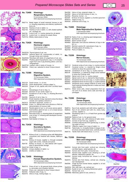
Prepared Microscope Slides Sets and Series 25<br />
Ma217d<br />
Ma2574d<br />
No. 72400<br />
Histology:<br />
Respiratory System,<br />
6 microscope slides<br />
With depictured accompanying brochure<br />
Ma211e Nasal region of small mammal (mouse or rat),<br />
t.s. showing respiratory and olfactory epithelium,<br />
bone etc.<br />
Ma215d Trachea of cat or rabbit, l.s.<br />
Ma214d Trachea of cat or rabbit, t.s. with ciliated epithelium,<br />
cartilage etc.<br />
Ma216c Lung of cat, t.s. routine stained for all details<br />
Ma217d Lung of cat, t.s. stained for elastic fibres<br />
Pa4101e Miliary tuberculosis of lung<br />
No. 72420<br />
Histology:<br />
Hormone organs<br />
6 microscope slides<br />
With depictured accompanying brochure<br />
Ma438d<br />
Ma439d<br />
Ma440e<br />
Ma445f<br />
Ma451d<br />
Ma454d<br />
No. 72480<br />
Uterus of pig, pregnant stage, t.s.<br />
Uterus of rat with embryo in situ, t.s.<br />
Placenta, human , t.s.<br />
Embryo of mouse, sagittal l.s. of entire specimen<br />
Vagina of pig, t.s.<br />
Umbilical cord of pig, t.s.<br />
Histology:<br />
Male Reproductive System,<br />
7 microscope slides<br />
With depictured accompanying brochure<br />
Ma4613d Testis of rat, t.s. showing spermatogenesis<br />
Ma463d Epididymis of bull, t.s.<br />
Ma464d Sperm smear of bull<br />
Ma466d Spermatic cord (Ductus deferens) of pig or rabbit,<br />
t.s.<br />
Ma467d Seminal vesicle (Gl. vesiculosa) of pig, t.s.<br />
Ma468d Prostate gland of monkey, t.s.<br />
Ma470d Penis of rabbit, t.s.<br />
Ma439d<br />
Ma4613d<br />
Ma255e<br />
Ma312d<br />
Ma331c<br />
Ma343f<br />
Ma346d<br />
Ma357d<br />
Ma411d<br />
Ma423c<br />
Ma2523d Thyroid gland of cat, sec.<br />
Ma253d Adrenal gland (Gl. suprarenalis) of rabbit, t.s.<br />
through cortex and medulla<br />
Ma2543d Pancreas with islets of Langerhans of cat, sec.<br />
Ma255e Pituitary gland (hypophysis), sag. l.s. of complete<br />
organ from cow or pig showing adeno- and neurohypophysis<br />
Ma2574d Leydig’s cells in testis of mouse, t.s.<br />
Ma434d Ovary, sec. selected to show Corpus luteum<br />
No. 72380<br />
Histology:<br />
Digestive System,<br />
15 microscope slides<br />
With depictured accompanying brochure<br />
Ma312d Tooth human, t.s. of root<br />
Ma316e Tooth development, medium stage l.s.<br />
Ma323d Tongue of cat, papilla with thick cornified layer,<br />
l.s.<br />
Ma331c Oesophagus of cat or dog, t.s.<br />
Ma334d Stomach of cat, fundic region t.s.<br />
Ma337c Duodenum of cat or dog, t.s. showing Brunner’s<br />
glands<br />
Ma338c Jejunum of cat or dog, t.s.<br />
Ma343f Small intestine of dog, injected to show the blood<br />
vessels and capillary network t.s.<br />
Ma341d Vermiform appendix, human t.s.<br />
Ma346d Colon, t.s. stained with muci-carmine or PAS for<br />
demonstration of mucous cells<br />
Ma351d Parotid gland of cat, t.s. of a pure serous gland<br />
Ma352d Submaxillary gland of cat, t.s. of a mixed serous<br />
and mucous gland<br />
Ma354d Pancreas of pig, t.s. showing islets of Langerhans<br />
Ma357d Liver of pig, t.s. showing well developed connective<br />
tissue<br />
Ma3634c Gall bladder of sheep, t.s.<br />
No. 72430<br />
Histology:<br />
Excretory System,<br />
6 microscope slides<br />
With depictured accompanying brochure<br />
Ma411d Kidney of cat, t.s. showing cortex with Malpighian<br />
corpuscles and medulla with tubules, Mallory’s<br />
stain<br />
Ma413e Kidney of mouse, sagittal l.s. through complete<br />
organ with cortex, medulla and pelvis<br />
Ma415f Kidney of mouse, t.s. vital stained with trypanblue<br />
to demonstrate storage<br />
Ma4214d Ureter of pig, t.s.<br />
Ma422c Urinary bladder of rabbit, t.s.<br />
Ma423c Urethra of rabbit, t.s.<br />
No. 72450<br />
Ma431d<br />
Ma434d<br />
Ma435c<br />
Ma437d<br />
Histology:<br />
Female Reproductive System,<br />
10 microscope slides<br />
With depictured accompanying brochure<br />
Ovary of cat, t.s. for general study, shows primary,<br />
secondary and Graafian follicles<br />
Ovary, sec. selected to show Corpus luteum<br />
Fallopian tube of pig, t.s.<br />
Uterus of pig, resting stage, t.s.<br />
No. 72250<br />
Histology:<br />
Nerve tissues,<br />
10 microscope slides<br />
With depictured accompanying brochure<br />
Ma511d Cerebral cortex of cat or dog, t.s. routine stained<br />
Ma512f Cerebral cortex, t.s. stained by Golgi’s silver<br />
method to show the pyramid cells<br />
Ma514d Cerebellum of cat or dog, t.s. routine stained<br />
Ma515f Cerebellum, t.s. stained by Golgi’s silver method<br />
to show the Purkinje cells<br />
Ma526d Spinal cord of cat, t.s. routine stained<br />
Ma527e Spinal cord of cat, t.s. stained for Nissl bodies<br />
Ma544c Peripheral nerve of cow or pig, l.s. routine stained<br />
Ma545c Peripheral nerve of cow or pig, t.s. routine stained<br />
Ma547e Peripheral nerve, teased material of osmic acid<br />
fixed material showing Ranvier’s nodes and medullary<br />
sheaths<br />
Ma551e Motor nerve cells, smear preparation from spinal<br />
cord of ox shows nerve cells and their appendages<br />
No. 72280<br />
Histology:<br />
Sense Organs,<br />
10 microscope slides<br />
With depictured accompanying brochure<br />
Ma601e Eye of cat, posterior part with retina, sagittal l.s.<br />
Ma602e Eye of cat, anterior part with iris, ciliary body,<br />
cornea, sagittal l.s.<br />
Ma608e Developing eyes in t.s. of head from guinea pig<br />
embryo<br />
Ma6034d Retina of cat, t.s. for general study<br />
Ma606f Retina of pig, sec. with entrance of optic nerve<br />
Ma607d Cornea of eye from pig, sagittal l.s.<br />
Ma609e Cochlea (internal ear) from guinea pig, l.s. showing<br />
organ of Corti<br />
Ma612d Olfactory region from nose of rabbit, t.s.<br />
Ma614e Taste buds, t.s. of papilla foliata in tongue of rabbit<br />
shows abundant taste buds, carefully stained<br />
Ma617e Tactile hairs with blood sinus, l.s. or t.s.<br />
No. 72350<br />
Histology:<br />
Skin and integument<br />
10 microscope slides<br />
With depictured accompanying brochure<br />
Ma632d Human skin from palm, vertical sec. showing<br />
cornified layers, sweat glands, etc.<br />
Ma633d Human skin from palm, horizontal sec.<br />
Ma635d Human scalp, sagittal l.s.sec. showing l.s. of hair<br />
follicles, sebaceous glands, etc.<br />
Ma636d Human scalp, horizontal sec. shows t.s. of hair<br />
follicles<br />
Ma637d Human skin from foetus, vertical sec. showing<br />
hair development<br />
Ma638e Finger tip from human foetus, sagittal l.s. of nail<br />
development<br />
Ma6404c Skin with hairs, cat, vertical sec.<br />
Ma6402c Eyelid of cat, t.s. showing Meibomian gland<br />
Ma647b Human hair, w.m.<br />
Ma6468d Mammary gland of cow, active t.s.<br />
Ma466d<br />
Ma467d<br />
Ma512f<br />
Ma514d<br />
Ma551e<br />
Ma608e<br />
Ma617e<br />
Ma635d<br />
Ma438d<br />
Ma637d


