

BIOLOGY - microscopia.info
BIOLOGY - microscopia.info
BIOLOGY - microscopia.info

Create successful ePaper yourself
Turn your PDF publications into a flip-book with our unique Google optimized e-Paper software.
Overhead Transparency Atlases<br />
99<br />
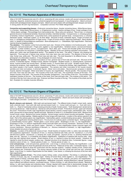
No. 8211E The Human Apparatus of Movement<br />
Atlas of 30 OHP Transparencies size 22 x 28 cm, comprising 66 color pictures, mostly with several component figures<br />
(drawings, diagrams, anatomical pictures, photomicrographs and macrographs, X-ray photographs). - Sketch and worksheets<br />
with semidiagrammatic designs and texts - Sketch and work-sheets with semidiagrammatic designs and texts -<br />
In strong plastic file with ring-mechanism. - Compilation and text: Prof. Walter Mergenthaler<br />
Connective and supporting tissues. - Embryonic connective tissue - Areolar connective tissue - White fibrous tissue,<br />
l.s. of tendon - Yellow elastic fibrous tissue, l.s. of ligamentum nuchae - Hyaline cartilage of frog - Costal cartilage of man<br />
- Yellow elastic cartilage - Fibrocartilage from intervertebral disc - Bone cells and canaliculi - Tibia of man, t.s. showing<br />
general structure: fundamental lamellae, Haversian lamellae, interstitial lamellae - Compact bone, t.s. showing systems<br />
of lamellae, medium magnification - Long hollow bone, entire epiphysis for general study - Compact bone, l.s. showing<br />
Haversian canals - Haversian system, t.s. for finer detail - Structure of bone, schematic figure - Finger of human embryo,<br />
l.s. cartilaginous predisposition of finger bones - Finger of human embryo, beginning ossification - Bone development,<br />
l.s. details of intracartilaginous ossification - Bone development, t.s. - Osteoblasts, high magnification - Red bone<br />
marrow showing megakaryocytes<br />
The skeleton. - The skeleton, entire front and entire back view - Division of the skeleton in its functional parts - Joints:<br />
hinge joint, ball-and-socket joint - Finger joint, sagittal l.s. low magnification - Vertebral column, cervical and thoracic<br />
vertebrae - Lumbar vertebra, sacrum, coccygeal bone - Skull, atlas, axis - Thorax and shoulder girdle, front and back<br />
views - Construction of a long bone, 3 schematic figures - Skeleton of the arm showing supination and pronation - The<br />
elbow joint, entire view and longitudinal section - The skeleton of the hand - The pelvis, 2 figures, one showing the<br />
ligaments - The knee joint, 4 figures: long. section, front view, back view, and menisci - The skeleton of the foot: side<br />
view, frontal view, ankle joint - The skull, front view and side view - The skull dissected in its different bones - X-ray<br />
photograph of a dislocation (luxation) - X-ray photograph of a bone fracture<br />
The muscular system. - The skeletal musculature of man, general view of front side and back side - Structure of the<br />
muscle, 4 schematic figures - Striated muscle, electron micrograph - Striated muscle, t.s. showing fascia, connective<br />
tissue, muscle bundles and muscle fibers - Striated muscle, l.s. muscle fibers and nuclei - Striated muscle fibers, l.s.<br />
showing the striations, high magnification - Striated muscle fibers, t.s. showing the fibrillae, high magnification - Capillary<br />
blood vessels in the muscle, injected preparation - The sensory and motor innervation of the muscle (muscle<br />
spindles and motor end plates), 4 schematic figures - Motor nerve end plates - Neuromuscular synapses in skeletal<br />
muscle, electron micrograph - Motor innervation of muscle, low magnification - Muscle spindle - The muscles of head<br />
and neck, front view and side view - The muscles of the trunk, front view - The superficial muscles of the back - The<br />
deeper muscles of the back - The muscles of the shoulder (antagonism) - The muscles of the arm - The pronation and<br />
supination muscles of the arm - The muscles of the hand, front view and back view - The muscles of the pelvis - The<br />
muscles of the leg, front view and side view - Extensor and flexor muscles of the leg - The muscles of the shank and the<br />
foot - Example of a complex muscular efficiency.<br />
No. 8212 E The Human Organs of Digestion<br />
Atlas of 30 OHP Transparencies size 22 x 28 cm, comprising 77 color pictures, mostly with several component figures<br />
(drawings, diagrams, anatomical pictures, photomicro- and macrographs) - Sketch and work-sheets with semidiagrammatic<br />
designs and texts - Compilation and text: Prof. W. Mergenthaler<br />
Mouth, pharynx and stomach. - Milk teeth and permanent teeth - The different kinds of teeth: incisor tooth, canine<br />
tooth, premolar tooth - Gum with milk tooth and permanent tooth, l.s. - Incisor tooth and gum, l.s. - Gum with root of<br />
tooth, t.s. - Head of mammalian embryo showing primordia of teeth, frontal section - Tooth development: dental lamina<br />
and young dental sac - Older dental sac - Dental sac with primordium of tooth - Primordium of tooth, upper part showing<br />
the crown - Primordium of tooth, high magnification shows dentine, enamel, enamal organ, odontoblastic cells - Human<br />
tooth, ground thin to show enamel, dentine and pulp - Bacteria of caries in l.s. of diseased human tooth - Bacteria from<br />
human mouth, smear - Bacteria from human intestine - Human tongue, section shows muscles and papillae - Tongue of<br />
cat, sec. with cornified papillae - Wallate papilla of human tongue with taste buds - Location of the salivary glands in the<br />
head - Part of the salivary gland, low magnification - Submaxillary gland, a predominating serous gland - Submaxillary<br />
gland, high magnification showing detail of acini - The structure of a salivary gland, schematic figure - Sublingual gland,<br />
a predominating mucous gland - Parotid gland, a pure serous gland - Esophagus of man, t.s. low magnification -<br />
Esophagus of man, t.s. medium magnification shows muscular layers and mucous membrane - Stomach of man, sagittal<br />
l.s. shows cardiac, fundic and pyloric region - Stomach, l.s. medium magnification shows muscular layers and<br />
mucous membrane - Mucous membrane of stomach, t.s. - Mucous membrane of stomach, t.s. high magnification -<br />
shows detailed structures of gastric glands<br />
Intestine. - Location of the abdominal viscera of man - Small intestine of newborn child, t.s. entire view and detail view<br />
with suspensory ligamentum - Duodenum of man, l.s. showing intestinal wall, folds, and villi - Duodenum, l.s. of a fold<br />
with Brunner’s glands - Duodenum, l.s. showing villi, crypts, and glands - Jejunum of man, l.s. showing intestinal wall,<br />
folds, and villi - Jejunum, l.s. of intestinal villi medium magnification - Epithelium of intestine with mucous cells - Intestinal<br />
loop with injected blood vessels - Small intestine of cat, t.s. injected to show the blood vessels - Intestinal villi<br />
injected to show the blood vessels, surface view - Detailed structure of an intestinal villus, 3 schematic figures - Large<br />
intestine (colon) of man, l.s. - Tubular glands of colon, l.s. - Tubular glands of colon, t.s.<br />
Liver and pancreas. - Liver and pancreas, general view - Liver of pig, t.s. shows liver lobules, low magnification - Liver<br />
lobule, schematic figure to show the glandular structure of the liver - Trabecula of liver cells, 2 schematic figures - Liver<br />
lobule, schematic figures to show the construction and the vascular systems - Capillary vessels of liver, central veins<br />
and collecting vein, schematic figure - The venous vascular system of the liver; portal vein and liver vein, schematic<br />
figure - Liver of pig, t.s. medium magnification for finer details - Liver lobule, t.s. showing the structure of the liver cells,<br />
high magnification<br />
The excretory system of man. - The urinary organs: kidney, ureter, urinary bladder - The kidney, l.s. schematic figure<br />
- Kidney of mouse, sag. sec. of complete organ - Kidney of human fetus, entire sagittal l.s., low magnification - The<br />
blood vessels of kidney, schematic figure - Human kidney, l.s. shows cortex, medulla, and pelvis, low magnification -<br />
Human kidney, t.s. of cortex, medium magnification - Malpighian corpuscle, showing Bowman’s capsule, glomerular<br />
loop of afferent and efferent arteries, convoluted tubules - Cortex of kidney, l.s. with injected blood vessels - Medulla of<br />
kidney, l.s. with renal tubules and collecting tubes - Kidney, injected with trypane blue to demonstrate storage in the<br />
convoluted tubules - Nephron and glomerulus, 2 schematic figures - Ureter, t.s. - Urinary bladder, t.s. of the wall


