

BIOLOGY - microscopia.info
BIOLOGY - microscopia.info
BIOLOGY - microscopia.info

Create successful ePaper yourself
Turn your PDF publications into a flip-book with our unique Google optimized e-Paper software.
36<br />
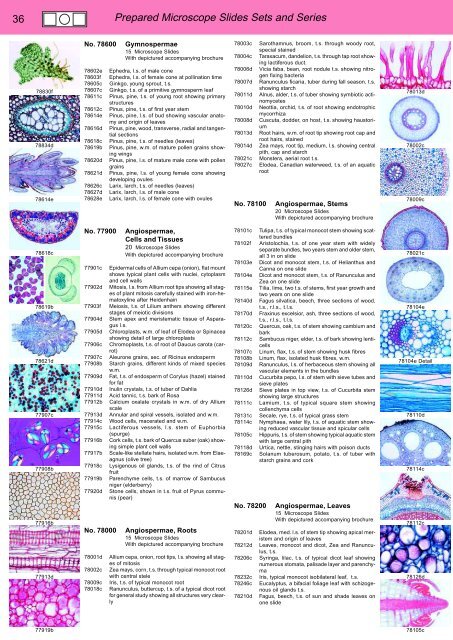
Prepared Microscope Slides Sets and Series<br />
78830f<br />
78834d<br />
78614e<br />
No. 78600<br />
78602e<br />
78603f<br />
78605c<br />
78607c<br />
78611c<br />
78612c<br />
78614e<br />
78616d<br />
78618c<br />
78619b<br />
78620d<br />
78621d<br />
78626c<br />
78627d<br />
78628e<br />
Gymnospermae<br />
15 Microscope Slides<br />
With depictured accompanying brochure<br />
Ephedra, l.s. of male cone<br />
Ephedra, l.s. of female cone at pollination time<br />
Ginkgo, young sprout, t.s.<br />
Ginkgo, t.s. of a primitive gymnosperm leaf<br />
Pinus, pine, t.s. of young root showing primary<br />
structures<br />
Pinus, pine, t.s. of first year stem<br />
Pinus, pine, l.s. of bud showing vascular anatomy<br />
and origin of leaves<br />
Pinus, pine, wood, transverse, radial and tangential<br />
sections<br />
Pinus, pine, t.s. of needles (leaves)<br />
Pinus, pine, w.m. of mature pollen grains showing<br />
wings<br />
Pinus, pine, l.s. of mature male cone with pollen<br />
grains<br />
Pinus, pine, l.s. of young female cone showing<br />
developing ovules<br />
Larix, larch, t.s. of needles (leaves)<br />
Larix, larch, l.s. of male cone<br />
Larix, larch, l.s. of female cone with ovules<br />
78003c<br />
78004c<br />
78006d<br />
78007d<br />
78011d<br />
78010d<br />
78008d<br />
78013d<br />
78014d<br />
78021c<br />
78027c<br />
No. 78100<br />
Sarothamnus, broom, t.s. through woody root,<br />
special stained<br />
Taraxacum, dandelion, t.s. through tap root showing<br />
lactiferous duct.<br />
Vicia faba, bean, root nodule t.s. showing nitrogen<br />
fixing bacteria<br />
Ranunculus ficaria, tuber during fall season, t.s.<br />
showing starch<br />
Alnus, alder, t.s. of tuber showing symbiotic actinomycetes<br />
Neottia, orchid, t.s. of root showing endotrophic<br />
mycorrhiza<br />
Cuscuta, dodder, on host, t.s. showing haustorium<br />
Root hairs, w.m. of root tip showing root cap and<br />
root hairs, stained<br />
Zea mays, root tip, medium, l.s. showing central<br />
pith, cap and starch<br />
Monstera, aerial root t.s.<br />
Elodea, Canadian waterweed, t.s. of an aquatic<br />
root<br />
Angiospermae, Stems<br />
20 Microscope Slides<br />
With depictured accompanying brochure<br />
78013d<br />
78002c<br />
78009c<br />
78618c<br />
78619b<br />
78621d<br />
77907c<br />
77908b<br />
77916b<br />
77913d<br />
No. 77900<br />
77901c<br />
77902d<br />
77903f<br />
77904d<br />
77905d<br />
77906c<br />
77907c<br />
77908b<br />
77909d<br />
77910d<br />
77911d<br />
77912b<br />
77913d<br />
77914c<br />
77915c<br />
77916b<br />
77917b<br />
77918c<br />
77919b<br />
77920d<br />
No. 78000<br />
78001d<br />
78002c<br />
78009c<br />
78018c<br />
Angiospermae,<br />
Cells and Tissues<br />
20 Microscope Slides<br />
With depictured accompanying brochure<br />
Epidermal cells of Allium cepa (onion), flat mount<br />
shows typical plant cells with nuclei, cytoplasm<br />
and cell walls<br />
Mitosis, l.s. from Allium root tips showing all stages<br />
of plant mitosis carefully stained with iron-hematoxyline<br />
after Heidenhain<br />
Meiosis, t.s. of Lilium anthers showing different<br />
stages of meiotic divisions<br />
Stem apex and meristematic tissue of Asparagus<br />
l.s.<br />
Chloroplasts, w.m. of leaf of Elodea or Spinacea<br />
showing detail of large chloroplasts<br />
Chromoplasts, t.s. of root of Daucus carota (carrot)<br />
Aleurone grains, sec. of Ricinus endosperm<br />
Starch grains, different kinds of mixed species<br />
w.m.<br />
Fat, t.s. of endosperm of Corylus (hazel) stained<br />
for fat<br />
Inulin crystals, t.s. of tuber of Dahlia<br />
Acid tannic, t.s. bark of Rosa<br />
Calcium oxalate crystals in w.m. of dry Allium<br />
scale<br />
Annular and spiral vessels, isolated and w.m.<br />
Wood cells, macerated and w.m.<br />
Lactiferous vessels, l.s. stem of Euphorbia<br />
(spurge)<br />
Cork cells, t.s. bark of Quercus suber (oak) showing<br />
simple plant cell walls<br />
Scale-like stellate hairs, isolated w.m. from Elaeagnus<br />
(olive tree)<br />
Lysigenous oil glands, t.s. of the rind of Citrus<br />
fruit<br />
Parenchyme cells, t.s. of marrow of Sambucus<br />
niger (elderberry)<br />
Stone cells, shown in t.s. fruit of Pyrus communis<br />
(pear)<br />
Angiospermae, Roots<br />
15 Microscope Slides<br />
With depictured accompanying brochure<br />
Allium cepa, onion, root tips, l.s. showing all stages<br />
of mitosis<br />
Zea mays, corn, t.s. through typical monocot root<br />
with central stele<br />
Iris, t.s. of typical monocot root<br />
Ranunculus, buttercup, t.s. of a typical dicot root<br />
for general study showing all structures very clearly<br />
78101c<br />
78102f<br />
78103e<br />
78104e<br />
78115e<br />
78140d<br />
78170d<br />
78120c<br />
78112c<br />
78107c<br />
78108b<br />
78109d<br />
78110d<br />
78126d<br />
78111c<br />
78131c<br />
78114c<br />
78105c<br />
78118d<br />
78169c<br />
No. 78200<br />
78201d<br />
78212d<br />
78206c<br />
78232c<br />
78246c<br />
78210d<br />
Tulipa, t.s. of typical monocot stem showing scattered<br />
bundles<br />
Aristolochia, t.s. of one year stem with widely<br />
separate bundles, two years stem and older stem,<br />
all 3 in on slide<br />
Dicot and monocot stem, t.s. of Helianthus and<br />
Canna on one slide<br />
Dicot and monocot stem, t.s. of Ranunculus and<br />
Zea on one slide<br />
Tilia, lime, two t.s. of stems, first year growth and<br />
two years on one slide<br />
Fagus silvatica, beech, three sections of wood,<br />
t.s., r.l.s., t.l.s.<br />
Fraxinus excelsior, ash, three sections of wood,<br />
t.s., r.l.s., t.l.s.<br />
Quercus, oak, t.s. of stem showing cambium and<br />
bark<br />
Sambucus niger, elder, t.s. of bark showing lenticells<br />
Linum, flax, t.s. of stem showing husk fibres<br />
Linum, flax, isolated husk fibres, w.m.<br />
Ranunculus, l.s. of herbaceous stem showing all<br />
vascular elements in the bundles<br />
Cucurbita pepo, l.s. of stem with sieve tubes and<br />
sieve plates<br />
Sieve plates in top view, t.s. of Cucurbita stem<br />
showing large structures<br />
Lamium, t.s. of typical square stem showing<br />
collenchyma cells<br />
Secale, rye, t.s. of typical grass stem<br />
Nymphaea, water lily, t.s. of aquatic stem showing<br />
reduced vascular tissue and spicular cells<br />
Hippuris, t.s. of stem showing typical aquatic stem<br />
with large central pith<br />
Urtica, nettle, stinging hairs with poison ducts<br />
Solanum tuberosum, potato, t.s. of tuber with<br />
starch grains and cork<br />
Angiospermae, Leaves<br />
15 Microscope Slides<br />
With depictured accompanying brochure<br />
Elodea, med. l.s. of stem tip showing apical meristem<br />
and origin of leaves<br />
Leaves, monocot and dicot, Zea and Ranunculus,<br />
t.s.<br />
Syringa, lilac, t.s. of typical dicot leaf showing<br />
numerous stomata, palisade layer and parenchyma<br />
Iris, typical monocot isobilateral leaf, t.s.<br />
Eucalyptus, a bifacial foliage leaf with schizogenous<br />
oil glands t.s.<br />
Fagus, beech, t.s. of sun and shade leaves on<br />
one slide<br />
78021c<br />
78104e<br />
78104e Detail<br />
78110d<br />
78114c<br />
78112c<br />
78126d<br />
77919b<br />
78105c


