

BIOLOGY - microscopia.info
BIOLOGY - microscopia.info
BIOLOGY - microscopia.info

Create successful ePaper yourself
Turn your PDF publications into a flip-book with our unique Google optimized e-Paper software.
Overhead Transparency Atlases<br />
117<br />
Vermiform appendix, t.s. - Small intestine with injected blood vessels, t.s. - Digestive System: Pancreas, Liver and<br />
Salivary Glands - Human pancreas, t.s. - Human liver, t.s. - Liver of pig, t.s. - Liver lobule, t.s. with injected bile<br />
canaliculi and t.s. with injected blood vessels - Submaxillary gland, t.s. - Sublingual gland t.s. - Parotid gland, t.s. -<br />
Excretory System - Kidney of mouse, sagittal l.s. - Kidney of cat, t.s. - Human renal cortex and medulla l.s. - Malpighian<br />
corpuscle, - Human urinary bladder, t.s. - Human ureter, t.s. - Reproductive Organs: Female - Ovary of cat, t.s. - Egg<br />
development: Young and older primary follicle, secondary, early and mature Graafian follicle, germ hillock and egg,<br />
ruptured Graafian follicle - Fallopian tube with embedded oocyte, t.s. - Corpus luteum t.s. - Ciliated epithelium of the<br />
Fallopian tube t.s. - Uterus, secretory, menstrual post-menstrual and pregnant t.s. - Placenta - Umbilical cord (navel<br />
string), t.s. - Vagina t.s. - Reproductive Organs: Male - Testis of mammal, t.s. show all stages of spermatogenesis -<br />
Human testis, interstitial cells of Leydig, t.s. - Testis, germinal epithelium - Epididymis t.s. - Seminal vesicle, t.s. -<br />
Spermatic cord (Ductus deferens), t.s. - Prostate of young man, t.s. - Penis, t.s. - Sperm smear - Nervous System -<br />
Peripheral nerve, human sciatic nerve, t.s. low, medium and high magnification - Medullated nerve fibers l.s. - Ranvier’s<br />
node, l.s., electron micrograph - Spinal cord t.s. silvered - Spinal cord with motor nerve cells - Gray and white matter of<br />
spinal cord, t.s. - Nerve cells with Nissl’s granules - Motor nerve cell - Cerebral cortex, human, t.s. - Cerebellum, human,<br />
t.s. - Pyramid cells - Purkinje cells - Pseudounipolar neuron (T-cell) - Spinal cord, spinal and sympathetic ganglion -<br />
Organs of Sense: The Eye - Eye, mammal and human, sagittal l.s. - Cornea, iris, ciliary body, lens - Papilla of optic<br />
nerve l.s. - Optic nerve, t.s. - Retina, t.s. high magnification - Retina, graphic design - Organs of Sense: The Ear -<br />
Cochlea l.s. - Organ of Corti l.s. - Organs of Sense: Smell and Taste - Olfactory region, t.s. - Olfactory epithelium with<br />
sensory cilia, t.s. - Tongue of rabbit with papilla foliata and taste buds, t.s. - Taste buds, high magnification - Vallate<br />
papilla of the human tongue t.s. - Organs of Sense: Touch and Perception - Corpuscle of Eimer, tactile organ l.s. -<br />
Tactile hairs with blood sinus, t.s. and l.s. - Vater-Pacinian corpuscles t.s. - Grandry’s and Herbst’s tactile corpuscles -<br />
Touch corpuscles in human skin, t.s. - Meissner’s corpuscle from human finger, design - Krause’s corpuscle, cold<br />
receptor, design - Integument, Skin and Scalp - Human skin from palm, l.s. cornified epidermis, germinative zone,<br />
sweat glands - Nail development of human embryo, sagittal l.s. of finger tip - Human skin, zone of keratinization and<br />
germinative zone - Human skin negro, t.s. with pigmented cells - Human skin with sweat glands, t.s. - Human skin l.s.<br />
injected to show the blood vessels - Human scalp, vertical section showing l.s. of hair follicles - Hair papilla and germinative<br />
zone, t.s. - Hair shaft with arrector pili muscle and sebaceous gland l.s. - Hair follicles in human scalp t.s. -<br />
Sebaceous glands t.s. - Human scalp, injected to show the blood vessels - Mammary gland, human t.s.<br />
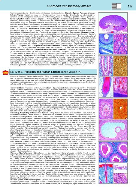
No. 8245 E Histology and Human Science (Short Version TA)<br />
Atlas of 30 Overhead-Transparencies size 22 x 28 cm, comprising over170 pictures (anatomical pictures, photomicroand<br />
macrographs, nature photographs, human photographs, electron micrographs, X-ray photographs, drawings, diagrams,<br />
tables, scenes, test data and results). With comprehensive interpretation text. Sketch and work-sheets with<br />
semidiagrammatic designs and texts - In strong plastic file with ring-mechanism. - Compilation and text: Dr. Karl-Heinrich<br />
Meyer B.S. and Johannes Lieder.<br />
Tissues and Skin: - Squamous epithelium, isolated cells - Squamous epithelium, color drawing and three dimensional<br />
design - Cuboidal epithelium in l.s. of kidney tubules - Columnar epithelium, human t.s. - Simple ciliated columnar<br />
epithelium, oviduct, t.s. - Pseudostratified ciliated columnar epithelium, trachea, t.s. - Areolar connective tissue, human<br />
- Areolar connective tissue, schematic color design - Adipose tissue, human, stained for fat - Adipose tissue, development,<br />
schematic color design - White fibrous tissue, tendon, human, l.s. - Yellow elastic connective tissue (Ligamentum<br />
nuchae), t.s. - Hyaline cartilage, human t.s. - Yellow elastic cartilage, human, sec. - White fibrous cartilage, human sec.<br />
- Compact bone, human tibia, t.s., low magnification - Compact bone, human t.s., high magnification showing Havers<br />
system - Spongy bone, human t.s., low magnification - Spongy bone, human t.s., high magnification for finer details -<br />
Bone development, l.s. of fetal finger - Striated (skeletal) muscle, human l.s., high magnification showing the striations<br />
- Striated muscle, t.s. of muscle bundle - Striated muscle t.s. high magnification for finer details - Smooth (involuntary)<br />
muscle, human l.s. - Smooth (involuntary) muscle, schematic color design - Heart (cardiac) muscle, human l.s. - Skin<br />
from finger tip, human, l.s. - Scalp, human, shows l.s. of hair follicles, - Scalp, shows t.s. of hair follicles, low magnification<br />
- Hair follicles from human scalp, high magnification.<br />
Circulatory, Respiratory and Endocrine System: - Artery, human, t.s. stained for elastic fibers - Vein, human, t.s.<br />
stained for elastic fibers - Aorta, human, t.s. - Artery and vein, human t.s., routine stained - Artery and vein, human t.s.,<br />
schematic color design - Blood smear, human, Giemsa stain - Blood smear, human, schematic color design - Frog blood<br />
smear, nucleated erythrocytes - Red blood cells (erythrocytes) of 12 species of animals for comparison, color design -<br />
Nasal region of small mammal, t.s. - Trachea, human t.s., low magnification - Trachea, human t.s., high magnification -<br />
Lung, human, t.s. - Lymph node, human t.s., general view, low magnification - Lymph node, human t.s., high magnification<br />
for fine details - Spleen, human t.s. - The vascular system of the human spleen, color diagram - Tonsil (Tonsilla<br />
palatina), human t.s. - Red bone marrow, human, smear - Thymus from human child, t.s. - Thyroid gland (Gl. thyreoidea),<br />
human t.s. - Adrenal gland (Gl. suprarenalis), human t.s. - Pituitary gland sag. l.s. of complete organ - Pituitary<br />
gland (Hypophysis), human t.s. - Location of pituitary gland and pineal body, sagittal l.s. of human head - Pineal body<br />
(Epiphysis), human t.s. - Islets of Langerhans in the pancreas, human, sec.<br />
Digestive System: - Lip, human foetus, t.s. - Tooth, human, t.s. of crown - Tooth, human, t.s. of root embedded in the<br />
yaw - Tooth development from human foetus, l.s. early stage - Tooth development from human foetus, l.s. later stage -<br />
Tongue, human, t.s. - Tongue of mouse, l.s. showing cornified papillae - Esophagus, human t.s., low magnification of the<br />
whole organ - Esophagus, human t.s. high magnification for fine detail - Stomach, fundic region, human t.s. - Duodenum,<br />
human t.s. - Jejunum, human t.s. - Vermiform appendix, human t.s. - Colon, human t.s., low magnification -<br />
Tubulous glands of the colon, detail, l.s. - Tubulous glands of the colon, detail, t.s. - Submaxillary gland (Gl. submandibularis),<br />
human t.s. - Sublingual gland (Gl. sublingualis), human t.s. - Parotid gland (Gl. parotis), human t.s. - Pancreas,<br />
human t.s. - Liver, human, t.s. - Liver, human, sec. staining of glycogen - Liver, of pig with liver lobules, t.s. low magnification<br />
- Liver lobule, t.s. with injected bile canaliculi - Vascular systems of a liver lobule, three dimensional color<br />
diagram - Gall bladder, human t.s.<br />
Urinary and Genital system: - Kidney, human l.s., low magnification - Kidney of mouse, sagittal l.s. through complete<br />
organ - Structure of kidney, color diagram - Human renal cortex, l.s., higher magnification - Human renal medulla, l.s. -<br />
Renal corpuscle (Malpighian corpuscle), high magnification - Kidney, sec. with injected blood vessels - Ureter, human<br />
t.s. - Urinary bladder, human t.s. - Ovary, mature, t.s. low magnification for general survey - Egg development: primary<br />
follicle - Egg development: secondary follicle - Egg development: mature Graafian follicle with germ hillock and egg cell<br />
- Egg development: Ruptured Graafian follicle after the oocyte has been discharged l.s. - Egg development: mature<br />
ovulated egg with corona radiata - Ovary with corpus luteum, human t.s. - Uterus, human, t.s. - Oviduct (fallopian tube),<br />
human, t.s. - Uterus of rat with embryo in situ, t.s. - Embryo of mouse, sagittal l.s. of entire specimen - Embryo of mouse,<br />
sagittal l.s. of head - Embryo of mouse, t.s. of thoracic region - Placenta, human t.s. - Structure and function of the<br />
placenta; diagram - Umbilical cord (navel string), human t.s. - Vagina, human t.s. - Mammary gland, human t.s. - Testis<br />
from human adult, mature stage t.s. - Testis t.s. stained to show all stages of spermatogenesis, high magnification -<br />
Interstitial cells of Leydig, in human testis t.s. - Testis, epididymis, spermatogenesis; color diagrams - Epididymis,<br />
human t.s. - Sperm smear of bull - Penis, t.s. - Seminal vesicle, t.s. - Prostate of young man, t.s. - Spermatic cord<br />
(Ductus deferens), human t.s.<br />
Nervous System and Sensory Organs: - Cerebral cortex, human, t.s. routine stained - Cerebral cortex, human, t.s.<br />
silvered for pyramidal cells - Cerebellum, human, t.s. routine stained - Cerebellum, human, t.s. silvered for Purkinje cells<br />
- Human brain, ventral view with cranial nerves - Brain of mouse, l.s. of the complete organ - Human spinal cord and


