

BIOLOGY - microscopia.info
BIOLOGY - microscopia.info
BIOLOGY - microscopia.info

You also want an ePaper? Increase the reach of your titles
YUMPU automatically turns print PDFs into web optimized ePapers that Google loves.
100<br />
Overhead Transparency Atlases<br />
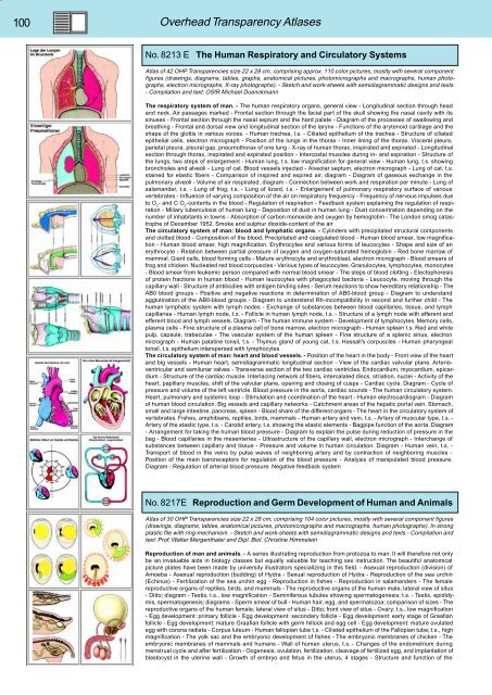
No. 8213 E The Human Respiratory and Circulatory Systems<br />
Atlas of 42 OHP Transparencies size 22 x 28 cm, comprising approx. 110 color pictures, mostly with several component<br />
figures (drawings, diagrams, tables, graphs, anatomical pictures, photomicrographs and macrographs, human photographs,<br />
electron micrographs, X-ray photographs). - Sketch and work-sheets with semidiagrammatic designs and texts<br />
- Compilation and text: OStR Michael Duenckmann<br />
The respiratory system of man. - The human respiratory organs, general view - Longitudinal section through head<br />
and neck. Air passages marked - Frontal section through the facial part of the skull showing the nasal cavity with its<br />
sinuses - Frontal section through the nasal septum and the hard palate - Diagram of the processes of swallowing and<br />
breathing - Frontal and dorsal view and longitudinal section of the larynx - Functions of the arytenoid cartilage and the<br />
shape of the glottis in various voices. - Human trachea, l.s. - Ciliated epithelium of the trachea - Structure of ciliated<br />
epithelial cells, electron micrograph - Position of the lungs in the thorax - Inner lining of the thorax. Visceral pleura,<br />
parietal pleura, pleural gap, pneumothorax of one lung - X-ray of human thorax, inspirated and expirated - Longitudinal<br />
section through thorax, inspirated and expirated position - Intercostal muscles during in- and expiration - Structure of<br />
the lungs, two steps of enlargement - Human lung, t.s. low magnification for general view - Human lung, t.s. showing<br />
bronchioles and alveoli - Lung of cat. Blood vessels injected - Alveolar septum, electron micrograph - Lung of cat, t.s.<br />
stained for elastic fibers - Comparison of inspired and expired air, diagram - Diagram of gaseous exchange in the<br />
pulmonary alveoli - Volume of air respirated, diagram - Connection between work and respiration per minute - Lung of<br />
salamander, t.s. - Lung of frog, t.s. - Lung of lizard, t.s. - Enlargement of pulmonary respiratory surface of various<br />
vertebrates - Influence of varying composition of the air on respiratory frequency - Frequency of nervous impulses due<br />
to O 2<br />
- and C O 2<br />
-contents in the blood - Regulation of respiration - Feedback system explaining the regulation of respiration<br />
- Miliary tuberculosis of human lung - Deposition of dust in human lung - Dust concentration depending on the<br />
number of inhabitants in towns - Absorption of carbon monoxide and oxygen by hemoglobin - The London smog catastrophe<br />
of December 1952. Smoke and sulphur dioxide-content of the air<br />
The circulatory system of man: blood and lymphatic organs. - Cylinders with precipitated structural components<br />
and clotted blood - Composition of the blood. Precipitated and coagulated blood - Human blood smear, low magnification<br />
- Human blood smear, high magnification. Erythrocytes and various forms of leucocytes - Shape and size of an<br />
erythrocyte - Relation between partial pressure of oxygen and oxygen-saturated hemoglobin - Red bone marrow of<br />
mammal. Giant cells, blood forming cells - Mature erythrocyte and erythroblast, electron micrograph - Blood smears of<br />
frog and chicken. Nucleated red blood corpuscles - Various types of leucocytes. Granulocytes, lymphocytes, monocytes<br />
- Blood smear from leukemic person compared with normal blood smear - The steps of blood clotting - Electophoresis<br />
of protein fractions in human blood - Human leucocytes with phagocyted bacteria - Leucocyte, moving through the<br />
capillary wall - Structure of antibodies with antigen binding sites - Serum reactions to show hereditary relationship - The<br />
AB0 blood groups - Positive and negative reactions in determination of AB0-blood group - Diagram to understand<br />
agglutination of the AB0-blood groups - Diagram to understand Rh-incompatibility in second and further child - The<br />
human lymphatic system with lymph nodes - Exchange of substances between blood capillaries, tissue, and lymph<br />
capillaries - Human lymph node, t.s. - Follicle in human lymph node, t.s. - Structure of a lymph node with afferent and<br />
efferent blood and lymph vessels. Diagram - The human immune system - Development of lymphocytes. Memory cells,<br />
plasma cells - Fine structure of a plasma cell of bone marrow, electron micrograph - Human spleen t.s. Red and white<br />
pulp, capsule, trabeculae - The vascular system of the human spleen - Fine structure of a splenic sinus, electron<br />
micrograph - Human palatine tonsil, t.s. - Thymus gland of young cat, t.s. Hassall’s corpuscles - Human pharyngeal<br />
tonsil, t.s. epithelium interspersed with lymphocytes<br />
The circulatory system of man: heart and blood vessels. - Position of the heart in the body - Front view of the heart<br />
and big vessels - Human heart, semidiagrammatic longitudinal section - View of the cardiac valvular plane. Arterioventricular<br />
and semilunar valves - Transverse section of the two cardiac ventricles. Endocardium, myocardium, epicardium<br />
- Structure of the cardiac muscle. Interlacing network of fibers, intercalated discs, striation, nuclei - Activity of the<br />
heart, papillary muscles, shift of the valvular plane, opening and closing of cusps - Cardiac cycle. Diagram - Cycle of<br />
pressure and volume of the left ventricle. Blood pressure in the aorta, cardiac sounds - The human circulatory system.<br />
Heart, pulmonary and systemic loop - Stimulation and coordination of the heart - Human electrocardiogram - Diagram<br />
of human blood circulation. Big vessels and capillary networks - Catchment areas of the hepatic portal vein. Stomach,<br />
small and large intestine, pancreas, spleen - Blood share of the different organs - The heart in the circulatory system of<br />
vertebrates. Fishes, amphibians, reptiles, birds, mammals - Human artery and vein, t.s. - Artery of muscular type, t.s. -<br />
Artery of the elastic type, t.s. - Carotid artery, t.s. showing the elastic elements - Bagpipe function of the aorta. Diagram<br />
- Arrangement for taking the human blood pressure - Diagram to explain the pulse during reduction of pressure in the<br />
bag - Blood capillaries in the mesenteries - Ultrastructure of the capillary wall, electron micrograph - Interchange of<br />
substances between capillary and tissue - Pressure and volume in human circulation. Diagram - Human vein, t.s. -<br />
Transport of blood in the veins by pulse waves of neighboring artery and by contraction of neighboring muscles -<br />
Position of the main baroreceptors for regulation of the blood pressure - Analysis of manipulated blood pressure.<br />
Diagram - Regulation of arterial blood pressure. Negative feedback system<br />
No. 8217E Reproduction and Germ Development of Human and Animals<br />
Atlas of 30 OHP Transparencies size 22 x 28 cm, comprising 104 color pictures, mostly with several component figures<br />
(drawings, diagrams, tables, anatomical pictures, photomicrographs and macrographs, human photographs). In strong<br />
plastic file with ring-mechanism. - Sketch and work-sheets with semidiagrammatic designs and texts - Compilation and<br />
text: Prof. Walter Mergenthaler and Dipl. Biol. Christine Himmelein<br />
Reproduction of man and animals. - A series illustrating reproduction from protozoa to man. It will therefore not only<br />
be an invaluable aids in biology classes but equally valuable for teaching sex instruction. The beautiful anatomical<br />
picture plates have been made by university illustrators specializing in this field. - Asexual reproduction (division) of<br />
Amoeba - Asexual reproduction (budding) of Hydra - Sexual reproduction of Hydra - Reproduction of the sea urchin<br />
(Echinus) - Fertilization of the sea urchin egg - Reproduction in fishes - Reproduction in salamanders - The female<br />
reproductive organs of reptiles, birds, and mammals - The reproductive organs of the human male; lateral view of situs<br />
- Ditto; diagram - Testis, t.s., low magnification - Seminiferous tubules showing spermatogenesis; t.s. - Testis, epididymis,<br />
spermatogenesis; diagrams - Sperm smear of bull - Human hair, egg, and spermatozoa; comparison of sizes - The<br />
reproductive organs of the human female; lateral view of situs - Ditto; front view of situs - Ovary; t.s., low magnification<br />
- Egg development: primary follicle - Egg development: secondary follicle - Egg development: early stage of Graafian<br />
follicle - Egg development: mature Graafian follicle with germ hillock and egg cell - Egg development: mature ovulated<br />
egg with corona radiata - Corpus luteum - Human fallopian tube t.s. - Ciliated epithelium of the Fallopian tube; t.s., high<br />
magnification - The yolk sac and the embryonic development of fishes - The embryonic membranes of chicken - The<br />
embryonic membranes of mammals and humans - Wall of human uterus, t.s. - Changes of the endometrium during<br />
menstrual cycle and after fertilization - Oogenesis, ovulation, fertilization, cleavage of fertilized egg, and implantation of<br />
blastocyst in the uterine wall - Growth of embryo and fetus in the uterus, 4 stages - Structure and function of the


