BIOLOGY - microscopia.info
BIOLOGY - microscopia.info
BIOLOGY - microscopia.info
You also want an ePaper? Increase the reach of your titles
YUMPU automatically turns print PDFs into web optimized ePapers that Google loves.
140<br />
Color Projection Slides and Photomicrographs 35 mm<br />
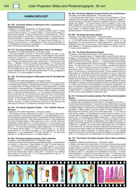
HUMAN <strong>BIOLOGY</strong><br />
No. 760. The Human System of Movement, Part I: Connective and<br />
Supporting Tissues.<br />
Compilation: Prof. Walter Mergenthaler. 19 Projection Slides<br />
1. Embryonic connective tissue 2. Loose (areolar) connective tissue 3. Dense<br />
connective tissue, tendon l.s. 4. Hyaline cartilage 5. Human costal cartilage 6.<br />
Yellow elastic cartilage 7. Fibrous cartilage from an intervertebral disc 8. Bone<br />
cells with processes 9. Human tibia, t.s. general structure 10. Bone tissue, t.s.<br />
systems of lamellae 11. Bone tissue, l.s. Haversian canals 12. Haversian system,<br />
t.s. 13. Fine structure of bone, diagram 14. Finger of human embryo l.s. 15.<br />
Phalanx of human embryo l.s. 16. Development of bone. Zone of calcification, l.s.<br />
17. Development of bone. Zone of ossification, t.s. 18. Osteoblasts (bone forming<br />
cells) 19. Red bone marrow with giant cells<br />
No. 770. The Human System of Movement, Part II: The Skeleton.<br />
Compilation: Prof. Walter Mergenthaler. 20 Projection Slides<br />
1. The human skeleton, front view 2. The human skeleton, rear view 3. Subdivision<br />
of the skeleton into its functional parts 4. Joints: diagram, hinge, ball-andsocket<br />
joint 5. Finger joint, l.s. 6. Spinal column, cervical and thoracal vertebrae<br />
7. Lumbar vertebrae, sacrum and coccyx 8. Articulations of the skull: skull, atlas,<br />
axis 9. Thorax and shoulder girdle, front and back view 10. Structure of a long<br />
bone 11. Skeleton of the arm, pronation and supination 12. The elbow joint,<br />
surface view and l.s. 13. The skeleton of the hand 14. The pelvic girdle with and<br />
without its ligaments 15. The knee joint: long. section, front view, back view, menisci<br />
16. The skeleton of the foot: side view, frontal view, ankle joint 17. The skull:<br />
anterior and lateral view 18. Skull with separated bones 19. X-ray photograph of<br />
a dislocation 20. X-ray photograph of a fracture<br />
No. 780. The Human System of Movement, Part III: The Muscular<br />
System.<br />
Compilation: Prof. Walter Mergenthaler. 23 Projection Slides<br />
1. Human body showing the skeletal muscles, front and rear views 2. The structure<br />
of a skeletal muscle, diagram 3. Skeletal (striated) muscle, t.s. 4. Skeletal muscle,<br />
l.s. low magnification 5. Skeletal muscle fibres, l.s. high magnification 6. Skeletal<br />
muscle fibres, t.s. high magnification 7. Capillaries and arteries of a skeletal muscle<br />
8. The sensory and motor innervation of a muscle 9. Motor end plates on muscle<br />
fibres 10. Muscle with muscle spindle, t.s. 11. The muscles of the head and the<br />
neck, front and side view 12. The muscles of the trunk, front view 13. The superficial<br />
muscles of the back 14. The deeper muscles of the back 15. The muscles of the<br />
shoulder 16. The muscles of the arm 17. Pronating and supinating muscles of the<br />
forearm 18. The muscles of the hand 19. The muscles of the pelvis 20. The<br />
muscles of the leg 21. Flexors and extensors of the leg 22. The muscles of the<br />
lower leg and of the foot 23. Example of a complex muscular action<br />
No. 810. The Human Digestive System, Part I: Mouth, Pharynx,<br />
Stomach.<br />
Compilation: Prof. Walter Mergenthaler. 24 Projection Slides<br />
1. The deciduous and the permanent set of teeth 2. The types of teeth: incisor,<br />
canine, premolar 3. Incisor tooth in the alveolus, l.s. 4. Jaw with dental root, t.s. 5.<br />
Head of pig embryo with dental primordia 6. Development of tooth: Dental lamina<br />
and early tooth primordium 7. Ditto: Older tooth primordium 8. Ditto: Dental sack<br />
with later tooth differentiation 9. Ditto: Apical part of crown 10. Ditto: Detail with<br />
ameloblasts, enamel, dentin etc. 11. Human tongue, t.s. 12. The position of the<br />
salivary glands in the head 13. Lobules of salivary gland 14. Human submaxillary<br />
gland, t.s. 15. Human submaxillary gland, higher magnification 16. The structure<br />
of the salivary glands, diagram 17. Human sublingual gland, t.s. 18. Human<br />
parotid gland, t.s. 19. Human esophagus, t.s., low magnification 20. Human<br />
esophagus, t.s., high magnification 21. Human stomach, l.s. 22. Wall of the<br />
stomach, t.s. 23. Gastric mucosa, l.s. 24. Gastric glands, l.s.<br />
No. 820. The Human Digestive System, Part II: Intestine.<br />
Compilation: Prof. Walter Mergenthaler. 16 Projection Slides<br />
1. Position and fixation of the human abdominal digestive organs. 2. Small intestine<br />
of a newborn, t.s. total view 3. Small intestine, t.s., detail 4. Human duodenum,<br />
l.s. 5. Duodenal fold, l.s. 6. Duodenal wall, l.s. 7. Human jejunum, l.s. 8. Human<br />
jejunum, l.s. with villi 9. Intestinal epithelium with goblet cells 10. Intestinal loop<br />
with blood vessels 11. Small intestine with injected blood vessels, t.s. 12. Intestinal<br />
villi with injected blood vessels, surface view 13. Structure of an intestinal<br />
villus, diagram 14. Human colon, l.s. 15. Tubulous glands of the colon, l.s. 16.<br />
Tubulous glands of the colon, t.s.<br />
No. 830. The Human Digestive System, Part III: Liver and Pancreas.<br />
Compilation: Prof. Walter Mergenthaler. 14 Projection Slides<br />
1. Liver and pancreas, surface view 2. Liver of pig, t.s. low magnification 3. General<br />
structure of a liver lobule, diagram 4. Structure of a hepatic cord, diagram 5.<br />
Vascular systems of a liver lobule, diagram 6. Capillaries of the liver, central vein,<br />
hepatic vein, diagram 7. The venous system of the liver, diagram 8. Liver of pig,<br />
higher magnification 9. Liver lobule, t.s. showing arrangement of the hepatic cords<br />
10. Liver lobule, t.s. with injected bile canaliculi 11. Liver lobule, t.s. with injected<br />
blood vessels 12. Glisson’s triangle; vein, artery, bile duct 13. Liver cells with<br />
glycogen granules 14. Human pancreas, t.s.<br />
No. 840. The Human Excretory System<br />
Compilation: Prof. Walter Mergenthaler. 12 Projection Slides<br />
1. The urinary organs: situs 2. Kidney, l.s., diagram 3. Kidney from human embryo,<br />
l.s. 4. The blood vessels of the kidney, diagram 5. Human kidney, l.s., low<br />
magnification 6. Human renal cortex, l.s., higher magnification 7. Renal corpuscle<br />
(Malpighian corpuscle) 8. Renal cortex, l.s. with injected blood vessels 9. Human<br />
renal medulla, l.s. 10. Nephron and glomerulus, diagram 11. Human ureter, t.s.<br />
12. Human urinary bladder, t.s.<br />
No. 743. The Human Respiratory System.<br />
Compilation: OStR Michael Duenckmann. 38 Projection Slides<br />
1. The human respiratory organs 2. Sagittal section through head and neck, air<br />
passages 3. Head with nasal cavities 4. Nasal septum and hard palate of human<br />
5. Swallowing and breathing 6. The larynx 7. Function of arytenoid cartilages,<br />
glottis and vocal cords 8. Human trachea, l.s. 9. Ciliary epithelium of the trachea,<br />
detail 10. Ciliated epithelial cells, electron micrograph 11. Position of lungs in the<br />
thorax 12. Inner lining of thorax 13. X-ray of thorax, inspirated and expirated<br />
position 14. Thorax showing inspiration and expiration, l.s. 15. Function of<br />
intercostal muscles 16. Detailed structure of the lung 17. Human pulmonary<br />
tissue 18. Human lung, t.s. bronchioles and alveoli 19. Lung, injected to show the<br />
blood vessels 20. The alveolar wall, electron micrograph 21. Lung, t.s. stained for<br />
elastic fibres 22. Comparison of inspired and expired air 23. Diagram of gaseous<br />
exchange in the alveoli 24. Volume of air respirated, diagram 25. Connection<br />
between work and respiration 26. Lung of salamander t.s. 27. Lung of frog t.s. 28.<br />
Lung of lizard t.s. 29. Enlargement of pulmonary respiratory surface of various<br />
vertebrates 30. Influence of varying composition of the air on respiratory frequency<br />
31. Position and function of the carotid bodies 32. Regulation of respiration, diagram<br />
33. Feedback system of the regulation of respiration 34. Miliary tuberculosis<br />
in human lung t.s. 35. Deposition of dust in human lung 36. Dust concentration<br />
in different towns 37. Absorption of carbon monoxide and oxygen by hemoglobin<br />
38. The London smog catastrophe of December 1952<br />
No. 747. The Human Circulatory System, Part I: Blood and Lymphatic<br />
Organs.<br />
Compilation: OStR Michael Duenckmann. 35 Projection Slides<br />
1. Cylinders with precipitated and clotted blood 2. Composition of the blood 3.<br />
Human blood smear, general view 4. Human blood smear, detail of blood corpuscles<br />
5. Shape and size of an erythrocyte 6. Pressure of oxygen and oxygen-saturated<br />
hemoglobin, diagram 7. Red bone marrow 8. Erythrocyte and erythroblast, electron<br />
micrograph 9. Blood smears of frog and chicken 10. Types of leucocytes 11.<br />
Blood smear from leukemic person and normal blood smear 12. The steps of<br />
blood clotting, diagram 13. Electophoresis of protein fractions in blood 14. Human<br />
leucocytes with phagocyted bacteria 15. Leucocyte, moving through the<br />
capillary wall 16. Antibodies with antigen binding sites 17. Serum reactions to<br />
show relationship 18. The AB0 blood groups 19. Positive and negative reactions<br />
of AB0-blood groups 20. Diagram of agglutination of the AB0-blood groups 21.<br />
Diagram of Rh-incompatibility 22. The human lymphatic system 23. Exchange of<br />
substances in capillaries 24. Human lymph node, general view 25. Follicle in<br />
human lymph node 26. Structure of a lymph node blood and lymph vessels 27.<br />
Human immune system, diagram 28. Development of lymphocytes. Memory cells<br />
29. Plasma cell, electron micrograph 30. Human spleen, t.s. 31. The vascular<br />
system of human spleen 32. Splenic sinus, electron micrograph 33. Human palatine<br />
tonsil, t.s. 34. Thymus gland, t.s. Hassall bodies 35. Human pharyngeal tonsil, t.s.<br />
No. 751. The Human Circulatory System, Part II: Heart and Blood<br />
Vessels.<br />
Compilation: OStR Michael Duenckmann. 32 Projection Slides<br />
1. Position of the heart in the human body 2. Front view of heart and big vessels<br />
3. Human heart, l.s. 4. View of the cardiac valvular plane 5. Transection of the two<br />
cardiac ventricles 6. Structure of the cardiac muscle l.s. 7. Activity of the heart 8.<br />
Cardiac cycle. Diagram 9. Pressure and volume of the left ventricle 10. Human<br />
circulatory system 11. Stimulation and coordination of the heart 12. Human<br />
electrocardiogram 13. Diagram of human blood circulation 14. Catchment areas<br />
of the portal vein 15. Blood share of the different organs 16. Heart and blood<br />
circulation of vertebrates 17. Human artery and vein, t.s. low magnification 18.


