BIOLOGY - microscopia.info
BIOLOGY - microscopia.info
BIOLOGY - microscopia.info
You also want an ePaper? Increase the reach of your titles
YUMPU automatically turns print PDFs into web optimized ePapers that Google loves.
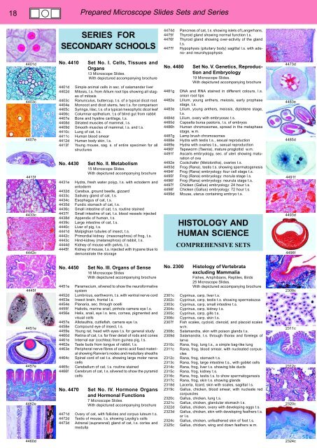
18<br />
Prepared Microscope Slides Sets and Series<br />
SERIES FOR<br />
SECONDARY SCHOOLS<br />
4474d<br />
4475f<br />
4476f<br />
4477f<br />
Pancreas of cat, t.s. showing islets of Langerhans,<br />
Thyroid gland showing normal function t.s.<br />
Thyroid gland showing over-activity of the gland<br />
t.s.<br />
Hypophysis (pituitary body) sagittal l.s. with adeno-<br />
and neurohypophysis<br />
4401d<br />
4403c<br />
4407e<br />
4413f<br />
4433c<br />
4442c<br />
No. 4410<br />
4401d<br />
4402d<br />
4403c<br />
4404e<br />
4405c<br />
4406c<br />
4407e<br />
4408d<br />
4409d<br />
4410c<br />
4411c<br />
4412d<br />
4413f<br />
No. 4430<br />
4431e<br />
4432d<br />
4433c<br />
4434c<br />
4435d<br />
4436c<br />
4437f<br />
4438d<br />
4439c<br />
4440c<br />
4441d<br />
4442c<br />
4443c<br />
4444d<br />
4445f<br />
Set No. I. Cells, Tissues and<br />
Organs<br />
13 Microscope Slides.<br />
With depictured accompanying brochure<br />
Simple animal cells in sec. of salamander liver<br />
Mitosis, l.s. from Allium root tips showing all stages<br />
of mitosis<br />
Ranunculus, buttercup, t.s. of a typical dicot root<br />
Monocot and dicot stems, two t.s. for comparison<br />
Syringa, lilac, t.s. of a typical mesophytic dicot leaf<br />
Columnar epithelium, t.s of blind gut from rabbit<br />
Bone and hyaline cartilage, t.s.<br />
Striated muscles of mammal, l.s.<br />
Smooth muscles of mammal, l.s. and t.s.<br />
Lung of cat, t.s.<br />
Human blood smear<br />
Human body skin, l.s.<br />
Young mouse, sag. s. of entire specimen for all<br />
structures<br />
Set No. II. Metabolism<br />
15 Microscope Slides.<br />
With depictured accompanying brochure<br />
Hydra, fresh water polyp, t.s. with ectoderm and<br />
entoderm<br />
Carabus, ground beetle, gizzard<br />
Salivary gland of cat, t.s.<br />
Esophagus of cat, t.s.<br />
Fundic stomach of cat, t.s.<br />
Small intestine of cat, t.s. routine stained<br />
Small intestine of cat, t.s. blood vessels injected<br />
Appendix of human, t.s.<br />
Large intestine of cat, t.s.<br />
Liver of pig, t.s.<br />
Malpighian tubules of insect, t.s.<br />
Primordial kidney (mesonephros) of frog, t.s.<br />
Hind-kidney (metanephros) of rabbit, t.s.<br />
Kidney of mouse with pelvis, l.s.<br />
Kidney of mouse, t.s. injected with trypane blue to<br />
demonstrate the storage<br />
No. 4480<br />
4481g<br />
4482e<br />
4483e<br />
4484d<br />
4485d<br />
4486h<br />
4487g<br />
4488e<br />
4489e<br />
4490f<br />
4491f<br />
4492e<br />
4493d<br />
4494f<br />
4495f<br />
4496f<br />
4497f<br />
4498f<br />
4499d<br />
Set No. V. Genetics, Reproduction<br />
and Embryology<br />
19 Microscope Slides.<br />
With depictured accompanying brochure<br />
DNA and RNA stained in different colours, l.s.<br />
onion root tips<br />
Lilium, young anthers, meiosis, early prophase<br />
stage, t.s.<br />
Lilium, young anthers, meiosis, diplotene stage,<br />
t.s.<br />
Lilium, ovary with embryosac t.s.<br />
Capsella bursa pastoris, l.s. of embryos<br />
Human chromosomes, spread in the metaphase<br />
stage, w.m.<br />
Lamp brush chromosomes<br />
Hydra with testis t.s., sexual reproduction<br />
Hydra with ovaries t.s., sexual reproduction<br />
Tapeworm (Taenia), mature proglottid w.m.<br />
Ascaris embryology, sec. of uteri showing maturation<br />
of ova<br />
Cockchafer (Melolontha), ovaries t.s.<br />
Frog (Rana), testis t.s. showing spermatogenesis<br />
Frog (Rana) embryology: four cell stage t.s.<br />
Frog (Rana) embryology: morula stage l.s.<br />
Frog (Rana) embryology: neurula stage t.s.<br />
Chicken (Gallus) embryology: 24 hour t.s.<br />
Chicken (Gallus) embryology: 72 hour t.s.<br />
Mouse, uterus containing embryo t.s.<br />
HISTOLOGY AND<br />
HUMAN SCIENCE<br />
COMPREHENSIVE SETS<br />
4473d<br />
4483e<br />
4485d<br />
4491f<br />
4493d<br />
4496f<br />
4445f<br />
4451e<br />
4457e<br />
4462e<br />
No. 4450<br />
4451e<br />
4452d<br />
4453e<br />
4454e<br />
4455f<br />
4456e<br />
4457e<br />
4458e<br />
4459e<br />
4460d<br />
4461e<br />
4462e<br />
4463e<br />
4464c<br />
4465c<br />
4466f<br />
No. 4470<br />
4471d<br />
4472d<br />
4473d<br />
Set No. III. Organs of Sense<br />
16 Microscope Slides<br />
With depictured accompanying brochure<br />
Paramecium, silvered to show the neuroformative<br />
system<br />
Lumbricus, earthworm, t.s. with ventral nerve cord<br />
Insect brain, frontal l.s.<br />
Planaria, sec. through ocelli<br />
Haliotis, marine snail, pinhole camera eye l.s.<br />
Helix, snail, eye l.s. lens, cornea, pigmented and<br />
visual cells<br />
Alloteuthis, cuttlefish, camera eye l.s.<br />
Compound eye of insect, l.s.<br />
Young rat, head with eyes t.s. for general study<br />
Retina of cat, t.s. for finer detail of rods and cones<br />
Internal ear (cochlea) from guinea pig, l.s.<br />
Taste buds from tongue of rabbit, t.s.<br />
Peripheral nerve fibres of osmic acid fixed material<br />
showing Ranvier’s nodes and medullary sheaths<br />
Spinal cord of cat t.s. showing large motor nerve<br />
cells<br />
Cerebellum of cat, t.s. routine stained<br />
Cerebrum of cat, t.s. silvered to show the pyramid<br />
cells<br />
Set No. IV. Hormone Organs<br />
and Hormonal Functions<br />
7 Microscope Slides.<br />
With depictured accompanying brochure<br />
Ovary of cat, with follicles and corpus luteum t.s.<br />
Testis of mouse, t.s. showing Leydig’s cells<br />
Adrenal (suprarenal) gland of cat, t.s. cortex and<br />
medulla<br />
No. 2300<br />
2301c<br />
2302c<br />
2303c<br />
2304c<br />
2305c<br />
2306c<br />
2307f<br />
2308c<br />
2309d<br />
2310c<br />
2311c<br />
2312c<br />
2313c<br />
2314c<br />
2315c<br />
2316c<br />
2317c<br />
2318d<br />
2319c<br />
2320c<br />
2321c<br />
2322d<br />
2323d<br />
2324c<br />
2325c<br />
Histology of Vertebrata<br />
excluding Mammalia<br />
Fishes, Amphibians, Reptiles, Birds<br />
25 Microscope Slides.<br />
With depictured accompanying brochure<br />
Cyprinus, carp, liver t.s.<br />
Cyprinus, carp, testis t.s. showing spermatozoa<br />
Cyprinus, carp, small intestine t.s.<br />
Cyprinus, carp, kidney t.s.<br />
Cyprinus, carp, gills t.s.<br />
Cyprinus, carp, skin t.s.<br />
Fish scales, cycloid, ctenoid, and placoid scales<br />
w.m.<br />
Salamandra, skin with poison glands t.s.<br />
Salamandra, t.s. through thorax and forelegs of<br />
larva<br />
Rana, frog, lung t.s., a simple bag-like lung<br />
Rana, frog, blood smear, with nucleated corpuscles<br />
Rana, frog, stomach t.s.<br />
Rana, frog, large intestine t.s., with goblet cells<br />
Rana, frog, liver t.s. showing bile ducts<br />
Rana, frog, kidney t.s.<br />
Rana, frog, testis t.s. to show spermatogenesis<br />
Rana, frog, skin t.s. showing glands<br />
Lacerta, lizard, skin with scales, sagittal l.s.<br />
Gallus, chicken, blood smear, with nucleate red<br />
corpuscles<br />
Gallus, chicken, lung t.s.<br />
Gallus, chicken, glandular stomach t.s.<br />
Gallus, chicken, ovary with developing eggs t.s.<br />
Gallus, chicken, skin with developing feathers t.s.<br />
or l.s.<br />
Gallus, chicken, unfeathered skin of foot t.s.<br />
Gallus, chicken, wing and down feathers w.m.<br />
2308c<br />
2311c<br />
2312c<br />
2320c<br />
4460d<br />
2324c


