

BIOLOGY - microscopia.info
BIOLOGY - microscopia.info
BIOLOGY - microscopia.info

You also want an ePaper? Increase the reach of your titles
YUMPU automatically turns print PDFs into web optimized ePapers that Google loves.
156<br />
Color Projection Slides and Photomicrographs 35 mm<br />
No. 3330. The Chicken Embryology (Gallus domesticus).<br />
20 Color Photomicrographs<br />
1. 6 hour, l.s. 2. 18 hour, w.m. 3. 24 hour, w.m. 4. 24 hour, t.s. primitive groove 5.<br />
24 hour, t.s. neural plate 6. 28 hour, w.m. 10 somites 7. 36 hour, t.s. posterior<br />
region of abdomen 8. 36 hour, t.s. of anterior region of abdomen 9. 36 hour, t.s.<br />
region of heart 10. 40 hour, w.m. 15 somites 11. 45 hour, l.s. 12. 48 hour, t.s. of<br />
abdomen 13. 50 hour, w.m. 14. 72 hour, w.m. blood vascular system injected 15.<br />
72 hour, t.s. posterior region of abdomen 16. 72 hour, t.s. region of head 17. 96<br />
hour, t.s. anterior region of abdomen 18. 96 hour, t.s. region of heart 19. 5 day,<br />
w.m. 20. 8 day, l.s.<br />
No. 3360. Development of Follicles in Mammalian Ovary.<br />
12 Color Photomicrographs<br />
1. Ovary t.s. for general study 2. Young primary follicles t.s. 3. Older primary<br />
follicle t.s. 4. Secondary follicle t.s. 5. Young Graafian follicle, l.s. 6. Older Graafian<br />
follicle, l.s. 7. Mature Graafian follicle, l.s. 8. Mature oocyte t.s. 9. Ruptured<br />
Graafian follicle l.s. 10. Fallopian tube with embedded oocyte, t.s. low magnification<br />
11. Ditto. t.s. detail 12. Ovary with Corpus luteum, t.s.<br />
No. 3340. The Eye Development in Vertebrates (Frog).<br />
10 Color Photomicrographs<br />
1. Early neurula, t.s. two pigmented grooves 2. Medium neurula, t.s. later stage 3.<br />
Later neurula, t.s. optic vesicles 4. Ditto. growing optic vesicles 5. Tail bud stage,<br />
t.s. formation of lens plate 6. Formation of the optic cup 7. Hatching larva, t.s.<br />
optic cup, optic stalk, brain 8. Fetal eye, l.s. entrance of mesenchyma and artery<br />
9. Eye of young tadpole, l.s. differentiation of lens and retina 10. Eye of older<br />
tadpole, l.s. of fully developed eye<br />
No. 3350. The Tooth Development. 10 Color Photomicrographs<br />
1. Early stage showing dental ridge l.s. 2. Young dental sac with bell-shaped<br />
enamel organ l.s. 3. Ditto. before formation of dentine and enamel 4. Later dental<br />
sac, formation of dentine l.s. 5. Ditto. formation of enamel l.s. 6. Formation of<br />
dentine and enamel, detail l.s. 7. Tooth shortly before dentition, detail l.s. 8. Gum<br />
with milk tooth and permanent tooth, l.s. 9. Gum with mature permanent tooth l.s.<br />
10. Gum with root of tooth, t.s.<br />
No. 725. Healing of Wounds and Regeneration. From the Wilhelm Roux<br />
Institute for Developmental Mechanics and Inheritance.<br />
Compilation: Dr. Hanns Koch. 18 Projection Slides<br />
1. Earthworm. Regeneration of the 4 anterior segments, one week after the<br />
operation 2. Ditto. after 4 weeks 3. Ditto. after 5 weeks 4. Frog tadpole. Regeneration<br />
of the tail after incision, after 2 weeks 5. Ditto. 4 weeks after the operation<br />
6. Salamander. Regeneration of the right foreleg, after 1 week 7. Ditto. after 2<br />
weeks 8. Ditto. after 3 weeks 9. Salamander. Regeneration of foreleg, diagrams<br />
10. Frog. Transplantation of a hindleg bud of a tadpole under the skin of the back<br />
of another tadpole. after 1 month 11. Salamander. Origin of the optic cup and<br />
lenses, diagrams 12. Ditto. Head l.s. 21 days after the cataract operation 13.<br />
Ditto. Left eye: retina deformed after 21 days 14. Ditto. Right eye: retina normal<br />
after 21 days 15. Ditto. Left eye, new lens, after 24 days 16. Ditto. Progressive<br />
formation of the new lens, after 30 days 17. Ditto. New lens free from the iris, after<br />
35 days 18. Ditto. New lens in the right place, end of regeneration after 50 days<br />
BOTANY - CRYPTOGAMS<br />
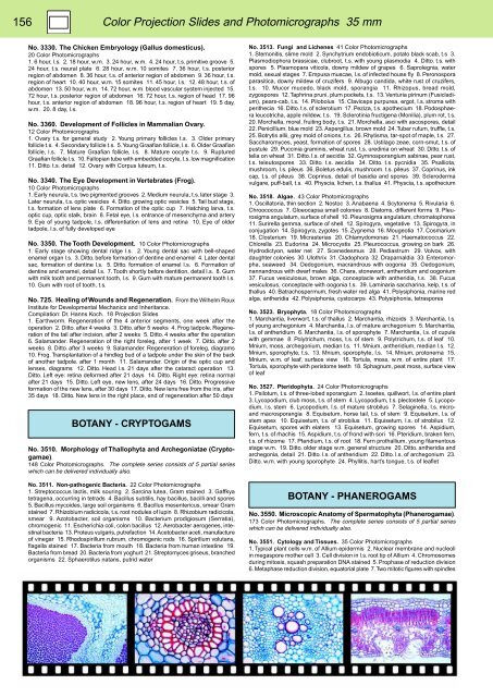
No. 3510. Morphology of Thallophyta and Archegoniatae (Cryptogamae).<br />
148 Color Photomicrographs. The complete series consists of 5 partial series<br />
which can be delivered individually also.<br />
No. 3511. Non-pathogenic Bacteria. 22 Color Photomicrographs<br />
1. Streptococcus lactis, milk souring 2. Sarcina lutea, Gram stained 3. Gaffkya<br />
tetragena, occurring in tetrads 4. Bacillus subtilis, hay bacillus, bacilli and spores<br />
5. Bacillus mycoides, large soil organisms 6. Bacillus mesentericus, smear Gram<br />
stained 7. Rhizobium radicicola, t.s. root nodules of lupin 8. Rhizobium radicicola,<br />
smear 9. Azotobacter, soil organisms 10. Bacterium prodigiosum (Serratia),<br />
chromogenic 11. Escherichia coli, colon bacillus 12. Aerobacter aerogenes, intestinal<br />
bacteria 13. Proteus vulgaris, putrefaction 14. Acetobacter aceti, manufacture<br />
of vinegar 15. Rhodospirillum rubrum, chromogenic rods 16. Spirillum volutans,<br />
flagella stained 17. Bacteria from mouth 18. Bacteria from human intestine 19.<br />
Bacteria from bread 20. Bacteria from yoghurt 21. Streptomyces griseus, branched<br />
organisms 22. Sphaerotilus natans, putrid water<br />
No. 3513. Fungi and Lichenes 41 Color Photomicrographs<br />
1. Stemonitis, slime mold 2. Synchytrium endobioticum, potato black scab, t.s 3.<br />
Plasmodiophora brassicae, clubroot, t.s. with young plasmodia 4. Ditto. t.s. with<br />
spores 5. Plasmopara viticola, downy mildew of grapes 6. Saprolegnia, water<br />
mold, sexual stages 7. Empusa muscae, l.s. of infected house fly 8. Peronospora<br />
parasitica, downy mildew of cruzifers 9. Albugo candida, white rust of cruzifers,<br />
t.s. 10. Mucor mucedo, black mold, sporangia 11. Rhizopus, bread mold,<br />
zygospores 12. Taphrina pruni, plum pockets, t.s. 13. Venturia pirinum (Fusicladium),<br />
pears-cab, t.s. 14. Pilobolus 15. Claviceps purpurea, ergot, l.s. stroma with<br />
perithecia 16. Ditto. t.s. of sclerotium 17. Peziza, t.s. apothecium 18. Podosphaera<br />
leucotricha, apple mildew, t.s. 19. Sclerotinia fructigena (Monilia), plum rot, t.s.<br />
20. Morchella, morel, fruiting body, t.s. 21. Morchella, asci with ascospores, detail<br />
22. Penicillium, blue mold 23. Aspergillus, brown mold 24. Tuber rufum, truffle, t.s.<br />
25. Botrytis allii, grey mold of onions, t.s. 26. Rhytisma, tar-spot of maple, t.s. 27.<br />
Saccharomyces, yeast, formation of spores 28. Ustilago zeae, corn-smut, t.s. of<br />
pustule 29. Puccinia graminis, wheat rust, t.s. uredinia on wheat 30. Ditto. t.s. of<br />
telia on wheat 31. Ditto. l.s. of aecidia 32. Gymnosporangium sabinae, pear rust,<br />
t.s. teleutospores 33. Ditto. t.s. aecidia 34. Ditto. t.s. pycnidia 35. Psalliota,<br />
mushroom, l.s. pileus 36. Boletus edulis, mushroom. t.s. pileus 37. Coprinus, ink<br />
cap, t.s. of pileus 38. Coprinus, detail of basidia and spores 39. Scleroderma<br />
vulgare, puff-ball, t.s. 40. Physcia, lichen, t.s. thallus 41. Physcia, t.s. apothecium<br />
No. 3518. Algae. 43 Color Photomicrographs<br />
1. Oscillatoria, thin section 2. Nostoc 3. Anabaena 4. Scytonema 5. Rivularia 6.<br />
Chroococcus 7. Gloeocapsa small colonies 8. Diatoms, different forms 9. Pleurosigma<br />
angulatum, surface of shell 10. Pleurosigma angulatum, chromatophores<br />
11. Surirella gemma, surface of shell 12. Spirogyra, vegetative 13. Spirogyra, in<br />
conjugation 14. Spirogyra, zygotes 15. Zygnema 16. Mougeotia 17. Cosmarium<br />
18. Closterium 19. Micrasterias 20. Chlamydomonas 21. Haematococcus 22.<br />
Chlorella 23. Eudorina 24. Microcystis 25. Pleurococcus, growing on bark 26.<br />
Hydrodictyon, water net 27. Scenedesmus 28. Pediastrum 29. Volvox, with<br />
daughter colonies 30. Ulothrix 31. Cladophora 32. Draparnaldia 33. Enteromorpha,<br />
seaweed 34. Oedogonium, macrandrous with oogonia 35. Oedogonium,<br />
nannandrous with dwarf males 36. Chara, stonewort, antheridium and oogonium<br />
37. Fucus vesiculosus, brown alga, conceptacle with antheridia, t.s. 38. Fucus<br />
vesiculosus, conceptacle with oogonia t.s. 39. Laminaria saccharina, kelp, t.s. of<br />
thallus 40. Batrachospermum, fresh water red alga 41. Polysiphonia, marine red<br />
alga, antheridia 42. Polysiphonia, cystocarps 43. Polysiphonia, tetraspores<br />
No. 3523. Bryophyta. 18 Color Photomicrographs<br />
1. Marchantia, liverwort, t.s. of thallus 2. Marchantia, rhizoids 3. Marchantia, l.s.<br />
of young archegonium 4. Marchantia, l.s. of mature archegonium 5. Marchantia,<br />
l.s. of antheridium 6. Marchantia, l.s. of sporophyte 7. Marchantia, l.s. of cupule<br />
with gemmae 8. Polytrichum, moss, t.s. of stem 9. Polytrichum, t.s. of leaf 10.<br />
Mnium, moss, archegonium, median l.s. 11. Mnium, antheridium, median l.s. 12.<br />
Mnium, sporophyte, t.s. 13. Mnium, sporophyte, l.s. 14. Mnium, protonema 15.<br />
Mnium, w.m. of leaf, surface view 16. Tortula, moss, w.m. of entire plant 17.<br />
Tortula, sporophyte with peristome teeth 18. Sphagnum, peat moss, surface view<br />
of leaf<br />
No. 3527. Pteridophyta. 24 Color Photomicrographs<br />
1. Psilotum, t.s. of three-lobed sporangium 2. Isoetes, quillwort, l.s. of entire plant<br />
3. Lycopodium, club moss, t.s. of stem 4. Lycopodium, t.s. plectostele 5. Lycopodium,<br />
l.s. stem 6. Lycopodium, l.s. of mature strobilus 7. Selaginella, l.s. microand<br />
macrosporangia 8. Equisetum, horse tail, t.s. of stem 9. Equisetum, l.s. of<br />
stem apex 10. Equisetum, t.s. of strobilus 11. Equisetum, l.s. of strobilus 12.<br />
Equisetum, spores with elaters 13. Equisetum, growing spores 14. Aspidium,<br />
fern, t.s. of rhachis 15. Aspidium, t.s. of frond with sori 16. Pteridium, braken fern,<br />
t.s. of rhizome 17. Pteridium, t.s. of root 18. Fern prothallium, young filamentous<br />
stage w.m. 19. Ditto. older stage w.m. general structure 20. Ditto. antheridia and<br />
archegonia, detail 21. Ditto. l.s. of antheridium 22. Ditto. l.s. of archegonium 23.<br />
Ditto. w.m. with young sporophyte 24. Phyllitis, hart’s tongue, t.s. of leaflet<br />
BOTANY - PHANEROGAMS<br />
No. 3550. Microscopic Anatomy of Spermatophyta (Phanerogamae).<br />
173 Color Photomicrographs. The complete series consists of 5 partial series<br />
which can be delivered individually also.<br />
No. 3551. Cytology and Tissues. 35 Color Photomicrographs<br />
1. Typical plant cells w.m. of Allium epidermis 2. Nuclear membrane and nucleoli<br />
in megaspore mother cell 3. Cell division in l.s. root tip of Allium 4. Chromosomes<br />
during mitosis, squash preparation DNA stained 5. Prophase of reduction division<br />
6. Metaphase reduction division, equatorial plate 7. Two mitotic figures with spindles


