

BIOLOGY - microscopia.info
BIOLOGY - microscopia.info
BIOLOGY - microscopia.info

Create successful ePaper yourself
Turn your PDF publications into a flip-book with our unique Google optimized e-Paper software.
120<br />
Overhead Transparency Atlases<br />
dex folliculorum, follicle mite of humans, w.m. and t.s. of human skin with parasites - Sarcoptes scabiei, itch mite, w.m.<br />
sec. through infected skin - Insecta: Lice and Bugs - Stomoxys, stable fly, piercing sucking mouth parts - Haematopinus<br />
suis, pig louse - Lipoptena cervi, louse fly - Phthirus pubis, pubic or crab louse, w.m. and egg (nit) attached to hair<br />
- Pediculus capitis, head louse - Cimex lectularius, bed bug, w.m. and graphic design - Insecta: Mosquitoes - Mosquito,<br />
Culex pipiens, life-cycle - Culex pipiens, mosquito, adult female and male w.m. - Anopheles, malaria mosquito, adult<br />
female and male, w.m. - Culex, mosquito, head and mouth parts of female and male, design - Culex, head and mouth<br />
parts of female and male w.m. - Anopheles sp., mouth parts of a female w.m. and male w.m. - Culex, eggs rafts w.m. -<br />
Culex, posterior end of larva w.m. - Culex, pupa w.m. - Culex, t.s. of mouth parts of female shows hypopharynx with the<br />
salivary duct - Anopheles, female sucking on human skin, photograph - Insecta: Fleas - Flea, habitus, anatomy and<br />
mouth parts, design - Pulex irritans, human flea, adult female and male w.m. - Xenopsylla cheopis, rat flea, carrier of the<br />
bubonic plague, adult female and male w.m. - Ctenocephalus canis, dog flea, adult female and male w.m. - Nosopsyllus<br />
fasciatus, rat flea, adult female and male w.m.<br />
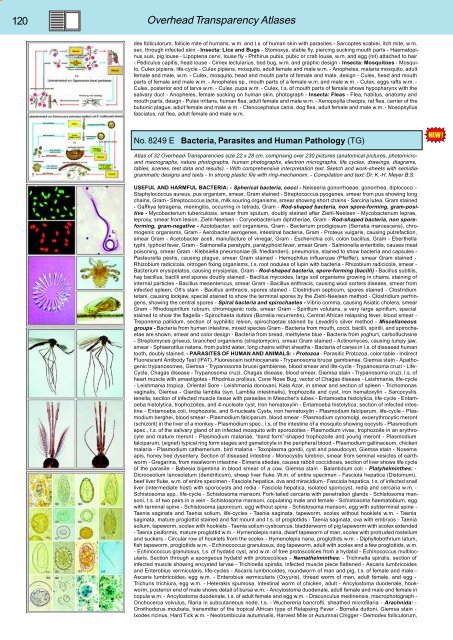
No. 8249 E Bacteria, Parasites and Human Pathology (TG)<br />
Atlas of 32 Overhead-Transparencies size 22 x 28 cm, comprising over 230 pictures (anatomical pictures, photomicroand<br />
macrographs, nature photographs, human photographs, electron micrographs, life cycles, drawings, diagrams,<br />
tables, scenes, test data and results). - With comprehensive interpretation text. Sketch and work-sheets with semidiagrammatic<br />
designs and texts - In strong plastic file with ring-mechanism. - Compilation and text: Dr. K.-H. Meyer B.S.<br />
USEFUL AND HARMFUL BACTERIA: - Spherical bacteria, cocci - Neisseria gonorrhoeae, gonorrhea, diplococci -<br />
Staphylococcus aureus, pus organism, smear, Gram stained - Streptococcus pyogenes, smear from pus showing long<br />
chains, Gram - Streptococcus lactis, milk souring organisms, smear showing short chains - Sarcina lutea, Gram stained<br />
- Gaffkya tetragena, meningitis, occurring in tetrads, Gram - Rod-shaped bacteria, non spore-forming, gram-positive<br />
- Mycobacterium tuberculosis, smear from sputum, doubly stained after Ziehl-Neelsen - Mycobacterium leprae,<br />
leprosy, smear from lesion, Ziehl-Neelsen - Corynebacterium diphtheriae, Gram - Rod-shaped bacteria, non sporeforming,<br />
gram-negative - Azotobacter, soil organisms, Gram - Bacterium prodigiosum (Serratia marcescens), chromogenic<br />
organisms, Gram - Aerobacter aerogenes, intestinal bacteria, Gram - Proteus vulgaris, causing putrefaction,<br />
smear Gram - Acetobacter aceti, manufacture of vinegar, Gram - Escherichia coli, colon bacillus, Gram - Eberthella<br />
typhi, typhoid fever, Gram - Salmonella paratyphi, paratyphoid fever, smear Gram - Salmonella enteritidis, causes meat<br />
poisoning, smear Gram - Klebsiella pneumoniae (B. friedlanderi), pneumonia, stained to show bacteria and capsules -<br />
Pasteurella pestis, causing plague, smear Gram stained - Hemophilus influenzae (Pfeiffer), smear Gram stained -<br />
Rhizobium radicicola, nitrogen fixing organisms, t.s. root nodules of lupin with bacteria - Rhizobium radicicola, smear -<br />
Bacterium erysipelatos, causing erysipelas, Gram - Rod-shaped bacteria, spore-forming (bacilli) - Bacillus subtilis,<br />
hay bacillus, bacilli and spores doubly stained - Bacillus mycoides, large soil organisms growing in chains, staining of<br />
internal particles - Bacillus mesentericus, smear Gram - Bacillus anthracis, causing wool sorters disease, smear from<br />
infected spleen, Olt’s stain - Bacillus anthracis, spores stained - Clostridium septicum, spores stained - Clostridium<br />
tetani, causing lockjaw, special stained to show the terminal spores by the Ziehl-Neelsen method - Clostridium perfringens,<br />
showing the central spores - Spiral bacteria and spirochaetes - Vibrio comma, causing Asiatic cholera, smear<br />
Gram - Rhodospirillum rubrum, chromogenic rods, smear Gram - Spirillum volutans, a very large spirillum, special<br />
stained to show the flagella - Spirochaeta duttoni (Borrelia recurrentis), Central African relapsing fever, blood smear -<br />
Treponema pallidum, section of syphilitic lesion, spirochaetae stained by Levaditi’s silver method - Miscellaneous<br />
groups - Bacteria from human intestine, mixed species Gram - Bacteria from mouth, cocci, bacilli, spirilli, and spirochaetae<br />
are shown, smear and color design - Bacteria from bread, methylene blue - Bacteria from yoghurt, carbolfuchsine<br />
- Streptomyces griseus, branched organisms (streptomycin), smear Gram stained - Actinomyces, causing lumpy jaw,<br />
smear - Sphaerotilus natans, from putrid water, long chains within sheaths - Bacteria of caries in l.s. of diseased human<br />
tooth, doubly stained. - PARASITES OF HUMAN AND ANIMALS: - Protozoa - Parasitic Protozoa, color table - Indirect<br />
Fluorescent Antibody Test (IFAT). Fluorescein isothiocyanate - Trypanosoma brucei gambiense, Giemsa stain - Apathogenic<br />
trypanosomes, Giemsa - Trypanosoma brucei gambiense, blood smear and life-cycle - Trypanosoma cruzi - Life-<br />
Cycle, Chagas disease - Trypanosoma cruzi, Chagas disease, blood smear, Giemsa stain - Trypanosoma cruzi, l.s. of<br />
heart muscle with amastigotes - Rhodnius prolixus, Cone Nose Bug, vector of Chagas disease - Leishmania, life-cycle<br />
- Leishmania tropica, Oriental Sore - Leishmania donovani, Kala Azar, in smear and section of spleen - Trichomonas<br />
vaginalis, Giemsa - Giardia lamblia (syn. Lamblia intestinalis), trophozoite and cyst, iron hematoxylin - Sarcocystis<br />
tenella, section of infected muscle tissue with parasites in Miescher’s tubes - Entamoeba histolytica, life-cycle - Entamoeba<br />
histolytica, trophozoites, and 4-nucleate cyst, Iron hematoxylin - Entamoeba histolytica, section of infected intestine<br />
- Entamoeba coli, trophozoite, and 8-nucleate Cysts, iron hematoxylin - Plasmodium falciparum, life-cycle - Plasmodium<br />
berghei, blood smear - Plasmodium falciparum, blood smear - Plasmodium cynomolgi, exoerythrocytic meront<br />
(schizont) in the liver of a monkey - Plasmodium spec., l.s. of the intestine of a mosquito showing oocysts - Plasmodium<br />
spec., t.s. of the salivary gland of an infected mosquito with sporozoites - Plasmodium vivax, trophozoite in an erythrocyte<br />
and mature meront - Plasmodium malariae, “band form”-shaped trophozoite and young meront - Plasmodium<br />
falciparum, (signet) typical ring form stages and gametocyte in the peripheral blood - Plasmodium gallinaceum, chicken<br />
malaria - Plasmodium cathemerium, bird malaria - Toxoplasma gondii, cyst and pseudocyst, Giemsa stain - Nosema<br />
apis, honey bee dysentery. Section of diseased intestine - Monocystis lumbrici, smear from seminal vesicles of earthworm<br />
- Gregarina, from mealworm intestine - Eimeria stiedae, causes rabbit coccidiosis, section of liver shows life cycle<br />
of the parasite - Babesia bigemina in blood smear of a cow, Giemsa stain - Balantidium coli - Platyhelminthes: -<br />
Dicroceolium lanceolatum (dendriticum), sheep liver fluke. W.m. of entire specimen - Fasciola hepatica (Distomum),<br />
beef liver fluke, w.m. of entire specimen - Fasciola hepatica, ova and miracidium - Fasciola hepatica, t.s. of infected snail<br />
liver (intermediate host) with sporocysts and redia - Fasciola hepatica, isolated sporocyst, redia and cercaria w.m. -<br />
Schistosoma spp., life-cycle - Schistosoma mansoni. Fork-tailed cercaria with penetration glands - Schistosoma mansoni,<br />
t.s. of two pairs in a vein - Schistosoma mansoni, copulating male and female - Schistosoma haematobium, egg<br />
with terminal spine - Schistosoma japonicum, egg without spine - Schistosoma mansoni, egg with subterminal spine -<br />
Taenia saginata and Taenia solium, life-cycles - Taenia saginata, tapeworm, scolex without hooklets w.m. - Taenia<br />
saginata, mature proglottid stained and flat mount and t.s. of proglottids - Taenia saginata, ova with embryos - Taenia<br />
solium, tapeworm, scolex with hooklets - Taenia solium cysticercus, bladderworm of pig tapeworm with scolex extended<br />
- Taenia pisiformis, mature proglottid w.m. - Hymenolepis nana, dwarf tapeworm of man, scolex with protruded rostellum<br />
and suckers - Circular row of hooklets from the scolex - Hymenolepis nana, proglottids w.m. - Diphyllobothrium latum,<br />
fish tapeworm, proglottids w.m. - Echinococcus granulosus, dog tapeworm, adult with scolex and a few proglottids, w.m.<br />
- Echinococcus granulosus, t.s. of hydatid cyst, and w.m. of free protoscolices from a hydatid - Echinococcus multilocularis.<br />
Section through a spongeous hydatid with protoscolices - Nemathelminthes: - Trichinella spiralis, section of<br />
infected muscle showing encysted larvae - Trichinella spiralis, infected muscle piece flattened - Ascaris lumbricoides<br />
and Enterobius vermicularis, life-cycles - Ascaris lumbricoides, roundworm of man and pig, t.s. of female and male -<br />
Ascaris lumbricoides, egg w.m. - Enterobius vermicularis (Oxyuris), thread worm of man, adult female, and egg -<br />
Trichuris trichiura, egg w.m. - Heterakis spumosa, intestinal worm of chicken, adult - Ancylostoma duodenale, hookworm,<br />
posterior end of male shows detail of bursa w.m. - Ancylostoma duodenale, adult female and male and female in<br />
copula w.m. - Ancylostoma duodenale, t.s. of adult female and egg w.m. - Dracunculus medinensis, macrophotograph -<br />
Onchocerca volvulus, filaria in subcutaneous node, t.s. - Wuchereria bancrofti, sheathed microfilaria - Arachnida: -<br />
Ornithodorus moubata, transmitter of the tropical African type of Relapsing Fever - Borrelia duttoni, Giemsa stain -<br />
Ixodes ricinus, Hard Tick w.m. - Neotrombicula autumnalis, Harvest Mite or Autumnal Chigger - Demodex folliculorum,


