

BIOLOGY - microscopia.info
BIOLOGY - microscopia.info
BIOLOGY - microscopia.info

You also want an ePaper? Increase the reach of your titles
YUMPU automatically turns print PDFs into web optimized ePapers that Google loves.
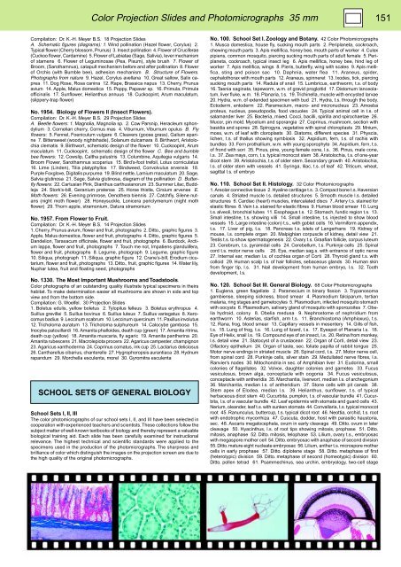
Color Projection Slides and Photomicrographs 35 mm 151<br />
Compilation: Dr. K.-H. Meyer B.S. 18 Projection Slides<br />
A. Schematic figures (diagrams): 1. Wind pollination (Hazel flower, Corylus) 2.<br />
Typical flower (Cherry blossom, Prunus) 3. Insect pollination 4. Flower of Cruciferae<br />
(Cuckoo flower, Cardamine) 5. Flower of Labiatae (Sage, Salivia), lever mechanism<br />
of stamens 6. Flower of Leguminosae (Pea, Pisum), style brush 7. Flower of<br />
Broom, (Sarothamnus), catapult mechanism before and after pollination 8. Flower<br />
of Orchis (with Bumble bee), adhesion mechanism B. Structure of Flowers.<br />
Photographs from nature 9. Hazel, Corylus avellana 10. Great sallow, Salix caprea<br />
11. Dog Rose, Rosa canina 12. Rape, Brassica napus 13. Cherry, Prunus<br />
avium 14. Apple, Malus domestica 15. Poppy, Papaver sp. 16. Primula, Primula<br />
officinalis 17. Sunflower, Helianthus annuus 18. Cuckoopint, Arum maculatum,<br />
(slippery-trap flower)<br />
No. 1954. Biology of Flowers II (Insect Flowers).<br />
Compilation: Dr. K.-H. Meyer B.S. 29 Projection Slides<br />
A. Beetle flowers: 1. Magnolia, Magnolia sp. 2. Cow Parsnip, Heracleum sphondylium<br />
3. Cornelian cherry, Cornus mas 4. Viburnum, Viburnum opulus B. Fly<br />
flowers: 5. Fennel, Foeniculum vulgare 6. Cleavers (goose grass), Galium aparine<br />
7. Bittersweet (woody nightshade), Solanum dulcamara 8. Birthwort, Aristolochia<br />
clematis 9. Birthwort, schematic design of the flower 10. Cuckoopint, Arum<br />
maculatum 11. Cuckoopint, schematic design of the flower C. Bee and bumble<br />
bee flowers: 12. Cowslip, Caltha palustris 13. Columbine, Aquilegia vulgaris 14.<br />
Broom Flower, Sarothamnus scoparius 15. Bird’s-foot trefoil, Lotus corniculatus<br />
16. Lime (Linden), Tilia platyphyllos 17. Bindweed, Convolvulus arvensis 18.<br />
Purple Foxglove, Digitalis purpurea 19. Blind nettle, Lamium maculatum 20. Sage,<br />
Salvia glutinosa 21. Sage, Salvia glutinosa, diagram of the pollination D. Butterfly<br />
flowers: 22. Cartusian Pink, Dianthus carthusianorum 23. Summer Lilac, Buddleja<br />
24. Stork’s-bill, Geranium pratense 25. Horse thistle, Cirsium arvense E.<br />
Moth flowers: 26. Evening primrose, Oenothera biennis 27. Catchfly, Silene nutans<br />
(night moth flower) 28. Honeysuckle, Lonicera periclymenum (night moth<br />
flower) 29. Thorn apple, stramonium, Datura stramonium<br />
No. 1957. From Flower to Fruit.<br />
Compilation: Dr. K.-H. Meyer B.S. 14 Projection Slides<br />
1. Cherry, Prunus avium, flower and fruit, photographs 2. Ditto., graphic figures 3.<br />
Apple, Malus domestica, flower and fruit, photographs 4. Ditto., graphic figures 5.<br />
Dandelion, Taraxacum officinale, flower and fruit, photographs 6. Burdock, Arctium<br />
lappa, flower and fruit, photographs 7. Touch me not, Impatiens glandulifera,<br />
flower and fruit, photographs 8. Legume, photograph 9. Legume, graphic figure<br />
10. Siliqua, photograph 11. Siliqua, graphic figure 12. Crane’s-bill, Erodium cicutarium,<br />
flower and fruit, photographs 13. Ditto., fruit, graphic figures 14. Water lily,<br />
Nuphar lutea, fruit and floating seed, photographs<br />
No. 1330. The Most Important Mushrooms and Toadstools.<br />
Color photographs of an outstanding quality illustrate typical specimens in theirs<br />
habitat. To make determination easier all mushrooms are shown in side and top<br />
view and from the bottom side.<br />
Compilation: G. Woelfel. 30 Projection Slides<br />
1. Boletus edulis, yellow boletus 2. Tylopilus felleus 3. Boletus erythropus 4.<br />
Suillus grevillei 5. Suillus bovinus 6. Suillus luteus 7. Suillus variegatus 8. Xerocomus<br />
badius 9. Leccinum scabrum 10. Leccinum quercinum 11. Paxillus involutus<br />
12. Tricholoma auratum 13. Tricholoma sulphureum 14. Calocybe gambosa 15.<br />
Inocybe patouillardi 16. Amanita phalloides, death cup (green) 17. Amanita ritrina,<br />
death cup (yellow) 18. Amanita muscaria, fly agaric 19. Amanita pantherina 20.<br />
Amanita rubescens 21. Macrolepiota procera 22. Agaricus campester, champignon<br />
23. Agaricus xanthoderma 24. Coprinus comatus, ink cup 25. Lactarius deliciosus<br />
26. Cantharellus cibarius, chanterelle 27. Hygrophoropsis aurantiaca 28. Hydnum<br />
rapandum 29. Morchella esculenta, morel 30. Gyromitra esculenta<br />
SCHOOL SETS OF GENERAL <strong>BIOLOGY</strong><br />
School Sets I, II, III<br />
The color photomicrographs of our school sets I, II, and III have been selected in<br />
cooperation with experienced teachers and scientists. These collections follow the<br />
subject matter of well-known textbooks of biology and thereby represent a valuable<br />
biological training aid. Each slide has been carefully examined for instructional<br />
relevance. The highest technical and scientific standards were applied to the<br />
specimens used in the production of the photomicrographs. The sharpness and<br />
brilliance of color which distinguish the images on the projection screen are due to<br />
the high quality of the original photomicrographs.<br />
No. 100. School Set I. Zoology and Botany. 42 Color Photomicrographs<br />
1. Musca domestica, house fly, sucking mouth parts 2. Periplaneta, cockroach,<br />
chewing mouth parts 3. Apis mellifica, honey bee, mouth parts of worker 4. Culex<br />
pipiens, common mosquito, piercing sucking mouth parts of adult female 5. Periplaneta,<br />
cockroach, typical insect leg 6. Apis mellifica, honey bee, hind leg of<br />
worker 7. Apis mellifica, wings 8. Pieris, butterfly, wing with scales 9. Apis mellifica,<br />
sting and poison sac 10. Daphnia, water flea 11. Araneus, spider,<br />
cephalothorax with mouth parts 12. Araneus, spinneret 13. Ixodes, tick, piercing<br />
sucking mouth parts 14. Radula of snail 15. Lumbricus, earthworm, t.s. of body<br />
16. Taenia saginata, tapeworm, w.m. of gravid proglottid 17. Distomum lanceolatum,<br />
liver fluke, w.m. 18. Planaria, t.s. 19. Trichinella, muscle with encysted larvae<br />
20. Hydra, w.m. of extended specimen with bud 21. Hydra, t.s. through the body.<br />
Ectoderm, entoderm 22. Paramecium, macro- and micronucleus 23. Amoeba<br />
proteus, nucleus, pseudopodia, food vacuoles 24. Typical animal cell in t.s. of<br />
salamander liver 25. Bacteria, mixed. Cocci, bacilli, spirilla and spirochaetae 26.<br />
Mucor, pin mold. Mycelium and sporangia 27. Coprinus, mushroom, section with<br />
basidia and spores 28. Spirogyra, vegetative with spiral chloroplasts 29. Mnium,<br />
moss, w.m. of leaf with cloroplasts 30. Diatoms, different species 31. Physcia,<br />
lichen, t.s. of thallus showing symbiosis 32. Aspidium, fern, t.s. of rachis with<br />
bundles 33. Fern prothallium, w.m. with young sporophyte 34. Aspidium, fern, t.s.<br />
of frond with sori 35. Pinus, pine, young female cone, l.s. 36. Pinus, male cone,<br />
l.s. 37. Zea mays, corn, t.s. typical monocot stem 38. Aristolochia, t.s. of one-year<br />
dicot stem 39. Aristolochia, t.s. of older stem. Secondary growth 40. Aristolochia,<br />
l.s. of older stem with vessels 41. Syringa, lilac, t.s. of leaf 42. Triticum, wheat,<br />
sagittal l.s. of embryo<br />
No. 110. School Set II. Histology. 32 Color Photomicrographs<br />
1. Areolar connective tissue 2. Hyaline cartilage t.s. 3. Compact bone t.s. Haversian<br />
canals 4. Striated muscle l.s. detailed structures 5. Smooth muscle l.s. detailed<br />
structures 6. Cardiac (heart) muscles, intercalated discs 7. Artery t.s. stained for<br />
elastic fibres 8. Vein t.s. stained for elastic fibres 9. Human blood smear 10. Lung<br />
t.s. alveoli, bronchial tubes 11. Esophagus t.s. 12. Stomach, fundic region t.s. 13.<br />
Small intestine, t.s. showing villi 14. Small intestine, t.s. injected to show blood<br />
vessels 15. Large intestine (colon) t.s., with goblet cells 16. Vermiform appendix,<br />
t.s. 17. Liver of pig, t.s. 18. Pancreas t.s. islets of Langerhans 19. Kidney of<br />
mouse, l.s. complete organ 20. Malpighian corpuscle of kidney, detail view 21.<br />
Testis t.s. to show spermatogenesis 22. Ovary t.s. Graafian follicle, corpus luteum<br />
23. Cerebrum, t.s. pyramidal cells 24. Cerebellum, t.s. Purkinje cells 25. Spinal<br />
cord t.s. motor nerve cells 26. Eye, median sag.s. with entrance of optic nerve<br />
27. Internal ear, median l.s. of cochlea organ of Corti 28. Thyroid gland t.s. with<br />
colloid 29. Human scalp l.s. of hair follicles, sebaceous glands 30. Human skin<br />
from finger tip, l.s. 31. Nail development from human embryo, l.s. 32. Tooth<br />
development, l.s.<br />
No. 120. School Set III. General Biology. 68 Color Photomicrographs<br />
1. Euglena, green flagellate 2. Paramecium in binary fission 3. Trypanosoma<br />
gambiense, sleeping sickness, blood smear 4. Plasmodium falciparum, tertian<br />
malaria, ring stages and gametocytes 5. Plasmodium, infected mosquito stomach<br />
with oocysts 6. Plasmodium, salivary gland of mosquito with sporozoites 7. Obelia<br />
hydroid, colony 8. Obelia medusa 9. Nephrostome of nephridium from<br />
earthworm 10. Asterias, starfish, arm t.s. 11. Branchiostoma (Amphioxus), t.s.<br />
12. Rana, frog, blood smear 13. Capillary vessels in mesentery 14. Gills of fish,<br />
t.s. 15. Lung of frog, t.s. 16. Lung of lizard, t.s. 17. Eyespot of Planaria l.s. 18.<br />
Eye of Helix, snail l.s. 19. Compound eye of an insect, l.s. 20. Retina from monkey,<br />
l.s. detail view 21. Statocyst of a crustacean 22. Organ of Corti, detail view 23.<br />
Olfactory epithelium 24. Organ of taste, sec. foliate papilla of rabbit tongue 25.<br />
Motor nerve endings in striated muscle 26. Spinal cord, t.s. 27. Motor nerve cell,<br />
from spinal cord 28. Purkinje cells, silver stain 29. Medullated nerve fibres, l.s.<br />
Ranvier’s nodes 30. Mitochondria in sec. of Amphibian liver 31. Eudorina, small<br />
colonies of flagellates 32. Volvox, daughter colonies and gametes 33. Fucus<br />
vesiculosus, brown alga, conceptacle with oogonia 34. Fucus vesiculosus,<br />
conceptacle with antheridia 35. Marchantia, liverwort, median l.s. of archegonium<br />
36. Marchantia, median l.s. of antheridium 37. Stone cells with pit canals 38.<br />
Stem apex of Elodea, median l.s. 39. Helianthus, sunflower, t.s. of typical<br />
herbaceous dicot stem 40. Cucurbita, pumpkin, t.s. of vascular bundle 41. Cucurbita,<br />
l.s. of a vascular bundle 42. Leaf epidermis with stomata and guard cells 43.<br />
Nerium, oleander, leaf t.s. with sunken stomata 44. Convallaria, t.s. typical monocot<br />
root 45. Ranunculus, buttercup, t.s. typical dicot root 46. Neottia, orchid, t.s. root<br />
with endotrophic mycorrhiza 47. Cuscuta, dodder, host with parasitic haustoria,<br />
sec. 48. Ascaris megalocephala, ovum in early cleavage 49. Ditto. ovum in later<br />
cleavage 50. Hyacinthus, l.s. of root tips showing mitosis, prophase 51. Ditto.<br />
mitosis, anaphase 52. Ditto. mitosis, telophase 53. Lilium, ovary t.s., embryosac<br />
with megaspore mother cell 54. Ditto. embryosac with anaphase of second division<br />
55. Ditto. mature eight nucleate embryosac 56. Lilium, anther t.s. microspore mother<br />
cells in early prophase 57. Ditto. diplotene stage 58. Ditto. metaphase of first<br />
(heterotypic) division 59. Ditto. metaphase of second (homeotypic) division 60.<br />
Ditto. pollen tetrad 61. Psammechinus, sea urchin, embryology, two-cell stage


