

BIOLOGY - microscopia.info
BIOLOGY - microscopia.info
BIOLOGY - microscopia.info

You also want an ePaper? Increase the reach of your titles
YUMPU automatically turns print PDFs into web optimized ePapers that Google loves.
Color Projection Slides and Photomicrographs 35 mm 137<br />
COLOR PROJECTION<br />
D518 Ranunculus,<br />
typical monocot ro<br />
t.s. of central stele<br />
D762 Motor nerve cells<br />
smear from spinal cord of cow with<br />
nerve cells and their processes<br />
SLIDES 35 mm<br />
PHOTOMICROGRAPHS<br />
Our new Color Slide Series are designed for modern teaching. They cover all requirements for teaching<br />
in secondary schools, high schools and colleges as well as in elementary schools. These visual aids will<br />
be invaluable in preparing students for the recognised examinations.<br />
The series of human biology are of great value in the training of nurses, medical technicians and for the<br />
students of physiotherapy and physical education. The study of the human body is the underlaying<br />
theme of the series which is usually the most important section of the school biology syllabus. This bias<br />
is also emphasised in the selection of anatomical and histological materials.<br />
The series of 35 mm slides consist of color pictures for histology and anatomy of the human body, color<br />
diagrams to illustrate the anatomical structures, color photomicrographs, human photographs, electron<br />
and scanning electron micrographs, X-ray photographs, drawings, diagrams, tables etc.<br />
The color pictures and diagrams have been prepared by university illustrators specialising in this field.<br />
In order to obtain maximum quality most of the slides delivered are ORIGINAL EXPOSURES, i.e. they<br />
are individually photographed from the specimen.<br />
The LIEDER series of projection slides will offer a complete range of slides covering all respects of<br />
school teaching in biology, physics and chemistry.<br />
LIEDER Color Photomicrographs for projection (on 35 mm film) are taken from selected prepared<br />
microscope slides. They immediately show, on the screen, the details of the specimen required for<br />
demonstration at the most suitable magnification. The student subsequently find it easier to locate the<br />
relevant part of a prepared slide under the microscope.<br />
In order to obtain maximum quality all the transparencies delivered are ORIGINAL EXPOSURES i.e.<br />
each LIEDER color photomicrograph is individually photographed from the specimen. Consequently,<br />
there is no loss of quality which could arise from a copying process.<br />
High quality photomicrographs can only be made from excellent, carefully selected microscope specimens.<br />
Every specimen used in the production of our photomicrographs has been either specially made<br />
or selected from many hundreds of preparations.<br />
Similar high standards must, of course, be applied to the selection and use of the photomicrographic<br />
apparatus. Our color photomicrographs are taken through microscopes with automatic cameras of the<br />
most advanced technique. These instruments are equipped with highly corrected optical systems, including<br />
flat field apochromatic objectives.<br />
LIEDER color photomicrographs are of high definition and clarity, coupled with color reproduction<br />
which has resulted in transparency slides of unsurpassed quality. They enable the maximum amount of<br />
<strong>info</strong>rmation to be illustrated in such a manner that it can be readily appreciated by the student.<br />
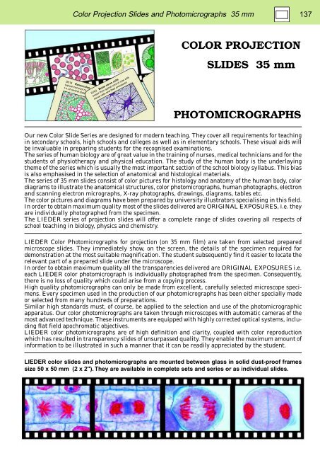
LIEDER color slides and photomicrographs are mounted between glass in solid dust-proof frames<br />
size 50 x 50 mm (2 x 2"). They are available in complete sets and series or as individual slides.


