

BIOLOGY - microscopia.info
BIOLOGY - microscopia.info
BIOLOGY - microscopia.info

You also want an ePaper? Increase the reach of your titles
YUMPU automatically turns print PDFs into web optimized ePapers that Google loves.
116<br />
Overhead Transparency Atlases<br />
mycelium and sporangia - Morchella, morel, t.s. of fruiting body with asci and spores - Claviceps purpurea, ergot,<br />
sclerotium t.s. - Saccharomyces, yeast, budding cells w.m. - Psalliota, mushroom, t.s. of pileus with basidia and<br />
spores - Puccinia graminis, wheat rust, uredinia on wheat leaf t.s. - Puccinia graminis, aecidia and pycnidia on<br />
barberry leaf t.s. - Physcia, lichen, thallus with symbiotic algae t.s. - Marchantia, liverwort, antheridia l.s. - Marchantia,<br />
archegonia l.s. - Moss stem with leaves w.m. - Sphagnum, peat moss, w.m. of leaf with chlorophyll-bearing and hyaline<br />
cells. - Fern prothallium, w.m. showing sex organs - Pteridium, bracken fern, rhizome t.s. - Aspidium, t.s. of leaf with<br />
sori, sporangia and spores - Equisetum, horse tail, strobilus with spores l.s.<br />
Botany, Phanerogams. - Allium cepa, onion, w.m. of epidermis shows simple plant cells - Root tip and root hairs -<br />
Zea mays, corn, monocot root t.s. - Ranunculus, buttercup, dicot root t.s. - Tilia, lime, woody dicot root t.s. - Dahlia, t.s.<br />
tuber with inuline crystals - Lupinus, lupin, root nodules with symbiotic bacteria t.s. - Elodea, waterweed, stem apex l.s.<br />
meristematic tissue and leaf origin - Zea mays, corn, monocot stem with scattered bundles t.s. - Helianthus, sunflower,<br />
herbaceous dicot stem t.s. - Pyrus, pear, t.s. of fruit with stone cells - Solanum tuberosum, potato, tuber with starch<br />
and cork cells t.s. - Elodea, waterweed, aquatic stem with primitive bundle t.s. - Triticum, wheat, t.s. of stem of a<br />
gramineous plant - Aristolochia, birthwort, one year stem t.s. - Aristolochia, older stem t.s. - Sambucus, elderberry,<br />
stem with lenticels t.s. - Tilia, lime, three sections of wood - Cucurbita, pumpkin, l.s. of stem with sieve tubes and<br />
vessels - Cucurbita, pumpkin, stem t.s. with sieve plates - Euphorbia, spurge, stem with lactiferous ducts l.s. - Salvia,<br />
sage, t.s. of a square stem with angular collenchyma - Tulipa, tulip, epidermis of leaf with stomata and guard cells w.m.<br />
- Iris, typical monocot leaf t.s. - Syringa, lilac, leaf t.s. - Fagus, beech, sun and shade leaves, two t.s. - Nerium,<br />
oleander, xerophytic leaf with sunken stomata, t.s. - Lilium, lily, anthers t.s. - Lilium, ovary t.s. showing arrangement of<br />
ovules - Taraxacum, dandelion, composite flower l.s. - Triticum, wheat, grain with embryo l.s. - Pinus, pine, three<br />
sections of wood - Pinus, pine, male cone with pollen l.s. - Pinus, female cone with ovules l.s. - Pinus, mature pollen<br />
grains with wings w.m.<br />
Cytology and Genetics. - Allium cepa, l.s. of root tips showing mitosis in all stages - Lilium, lily, t.s. of young anthers,<br />
meiotic stages of the pollen mother cells - Salamandra larva, sections with mitotic stages - Mitochondria, in thin sec.<br />
- Golgi apparatus, t.s. through spinal ganglion - Chloroplasts, in leaf of Elodea or Mnium, special stained - Aleurone<br />
grains, in sec. of Ricinus endosperm - Allium cepa, onion, w.m. of dry scale showing calcium oxalate crystals -<br />
Storage, section of liver or kidney, vital stained with trypan-blue to demonstrate storage - DNA in cell nuclei, by<br />
Feulgen staining technique - DNA and RNA, fixed and stained with methyl green and pyronine to show DNA and RNA<br />
in different colors - Giant chromosomes from the salivary gland of Chironomus. Individual genes and puffs can be<br />
observed - Human chromosomes, spread in the stage of metaphase, for counting chromosomes - Meiotic and mitotic<br />
stages in crayfish testis. Nuclear spindles - Maturation divisions in ova of Ascaris megalocephala - Cleavage<br />
stages in ova of Ascaris<br />
Embryology. - Chicken embryo, 48 hour, t.s. with neural tube and chorda - Sea-urchin development (Psammechinus<br />
miliaris), two cell, four cell and eight cell stages - Sea-urchin development (Psammechinus miliaris), morula, blastula<br />
and gastrula - Frog embryology (Rana), sec. trough the blastula stage showing the blastocoel - Frog embryology<br />
(Rana), sag. sec. through young larva in the tail bud stage, with primordia of organs<br />
Bacteria and Diseased Organs of Man. - Escherichia coli, bacteria from colon, probably pathogenic, smear Gram<br />
stained - Eberthella typhi, causing typhoid fever, smear Gram stained - Tuberculous lung of man, t.s. with miliary<br />
tuberculosis - Coal dust lung (Anthracosis) of man, t.s. (smoker’s lung) - Liver cirrhosis of man caused by alcohol<br />
abuse, t.s. showing degeneration of liver cells - Arteriosclerosis, t.s. of diseased coronary artery - Metastatic carcinoma<br />
(cancer) of human liver, t.s.<br />
Ecology and Environment. - Leaf (needle) of fir (Abies), two t.s. of leaves, healthy and damaged by environmental<br />
influences (acid rain) - Leaf of beech (Fagus), two t.s. of leaves, healthy and damaged by environmental influences<br />
(acid rain) - Bacteria from waste-water, smear with many typical forms.<br />
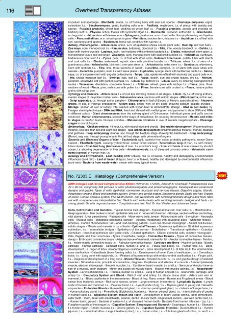
No. 72303 E Histology (Comprehensive Version)<br />
NEW enlarged and revised Comprehensive Edition (former no. 172303). Atlas of 41 Overhead-Transparencies size<br />
22 x 28 cm, comprising 228 pictures of color photomicrographs and photomacrographs, histological and anatomical<br />
designs and graphs. Types of cells. Epithelial, connective, muscular and nervous tissues. Digestive organs. Glands.<br />
Respiratory organs. Blood and lymphatic system. Urinary and genital organs. Endocrine glands. Scalp and hair. Organs<br />
of sense. Central nervous system. Plus NEW Sketch- and worksheets with semidiagrammatic designs and texts. Manual<br />
with comprehensive interpretation text. Sketch and work-sheets with semidiagrammatic designs and texts - In<br />
strong plastic file with ring-mechanism. - Compilation and text: Prof. Dr. Kurt Fiedler and Johannes Lieder<br />
Cells, Cell Division and Genetics - Typical Animal Cell, diagram - Typical animal cell, liver cells t.s. - Mitochondria -<br />
Golgi apparatus - Barr bodies in mouth epithelial cells and in nerve cell of woman - Storage, sections of liver and kidney,<br />
vital stained - Liver parenchyma - Pigment cells - Motor nerve cells, smear - Polynucleate cells - Syncytium - Neuroglia<br />
cells - Mucous cells - Metastatic carcinoma (cancer) - Ascaris, metaphase with equatorial plate - Whitefish mitosis -<br />
Amitosis - Human chromosomes, GTB-and RBA-banding pattern - Liver cell electron micrograph - Animal cell division,<br />
mitotic stages - Mitosis and meiosis in t.s. of testis - Epithelial Tissues. - Squamous epithelium - Stratified squamous<br />
epithelium, t.s. - Intercellular bridges - Epithelium of the cornea - Endothelium - Transitional epithelium - Cuboidal<br />
epithelium - Intestinal epithelium with goblet cells - Ciliated epithelium - Ciliated epithelial cells, electron micrograph -<br />
Cilia, flagella and their structures - Types of epithelia, design - Connective Tissues - Types of connective tissues,<br />
design - Embryonic connective tissue - Adipose tissue of mammal, stained for fat - Areolar connective tissue - Tendon,<br />
l.s. - Yellow elastic connective tissue t.s. - Reticular connective tissue - Cartilage and Bone - Hyaline cartilage - Elastic<br />
cartilage - Fibrous cartilage - Compact bone, human t.s, and l.s. - Fibula (calf-bone), t.s. - Human tibia, t.s. - Bone<br />
development, l.s. finger of fetus, intracartilaginous ossification - Bone development, t.s. of fetal skull, the intermembranous<br />
ossification - Osteoblasts (bone forming cells), t.s. - Development of bone. Zone of ossification, t.s. - Cancellous<br />
bone, t.s. - Long bone with epiphysis, l.s. - Phalanx of human embryo with endochondral ossification, l.s. - Finger joint,<br />
l.s. - Diagram of development of a long bone - Muscle Tissues - Striated muscle l.s., t.s. and graphic design of skeletal<br />
muscles - Striated muscle, principle of contraction, diagram - Capillaries and arteries of a muscle - Striated (skeletal)<br />
muscle, electron micrograph - Smooth muscles, l.s. - Cardiac or heart muscle, t.s. and l.s. - Sensory and motor innervation<br />
of a muscle, color diagram - Motor end plates on muscle fibers - Muscle with muscle spindle, t.s. - Respiratory<br />
System - Larynx of mammal, l.s. - Trachea, human t.s. and l.s. - Lung of human and cat, t.s. - Bronchiole, cartilage, and<br />
artery t.s. - Circulatory System and Blood - Wall of vein and artery, t.s. elastic tissue stain - Artery and vein of<br />
mammal, t.s. - Blood capillaries in the mesenteries - Blood of frog, Rana, smear - Human and Frog blood smear - Blood<br />
smear from leukemic person - Red bone marrow, smear - Large omentum (mesentery) - Lymphatic System - Lymph<br />
node of human and mammal, t.s. - Palatine tonsil, t.s. - Lymph node of pig, t.s. - Thymus gland of young cat, Hassall’s<br />
corpuscles - Endocrine Glands - Human thyroid gland, t.s. - Human parathyroid gland, t.s. - Islands of Langerhans, t.s.<br />
- Human pituitary gland, l.s. - Pineal body (Epiphysis), human t.s. - Human adrenal gland, t.s. - Interstitial cells of Leydig<br />
in human testis t.s. - Digestive System: Mouth and Teeth - Development of tooth: Dental lamina, tooth primordium,<br />
older tooth - Tooth, detail with ameloblasts, enamel, dentin - Incisor tooth, longitudinal section - Jaw with dental root, t.s.<br />
- Human tooth, ground - Bacteria of caries in l.s. of diseased human tooth - Bacteria from human intestine - Lip, t.s. -<br />
Fungiform papilla of the tongue t.s. - Digestive System: Esophagus and Stomach - Esophagus, human t.s. - Stomach<br />
t.s. fundic region - Gastric mucosa, l.s. - Gastric glands, l.s. - Digestive System: Intestine - Duodenal fold, l.s. - Human<br />
jejunum, l.s. - Intestinal villus - Large intestine (colon), t.s. - Human colon, l.s. - Tubulous glands of colon, l.s. and t.s. -


