Organohalogen concentrations and a gross and histologic ...
Organohalogen concentrations and a gross and histologic ...
Organohalogen concentrations and a gross and histologic ...

Create successful ePaper yourself
Turn your PDF publications into a flip-book with our unique Google optimized e-Paper software.
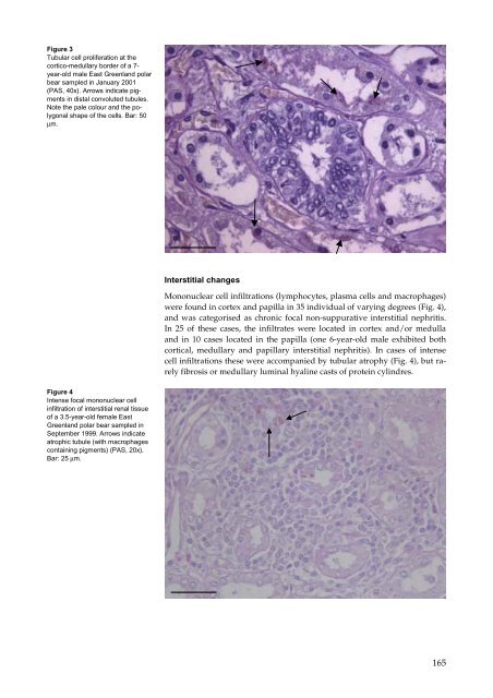
Figure 3<br />
Tubular cell proliferation at the<br />
cortico-medullary border of a 7year-old<br />
male East Greenl<strong>and</strong> polar<br />
bear sampled in January 2001<br />
(PAS, 40x). Arrows indicate pigments<br />
in distal convoluted tubules.<br />
Note the pale colour <strong>and</strong> the polygonal<br />
shape of the cells. Bar: 50<br />
µm.<br />
Figure 4<br />
Intense focal mononuclear cell<br />
infiltration of interstitial renal tissue<br />
of a 3.5-year-old female East<br />
Greenl<strong>and</strong> polar bear sampled in<br />
September 1999. Arrows indicate<br />
atrophic tubule (with macrophages<br />
containing pigments) (PAS, 20x).<br />
Bar: 25 µm.<br />
Interstitial changes<br />
Mononuclear cell infiltrations (lymphocytes, plasma cells <strong>and</strong> macrophages)<br />
were found in cortex <strong>and</strong> papilla in 35 individual of varying degrees (Fig. 4),<br />
<strong>and</strong> was categorised as chronic focal non-suppurative interstitial nephritis.<br />
In 25 of these cases, the infiltrates were located in cortex <strong>and</strong>/or medulla<br />
<strong>and</strong> in 10 cases located in the papilla (one 6-year-old male exhibited both<br />
cortical, medullary <strong>and</strong> papillary interstitial nephritis). In cases of intense<br />
cell infiltrations these were accompanied by tubular atrophy (Fig. 4), but rarely<br />
fibrosis or medullary luminal hyaline casts of protein cylindres.<br />
165