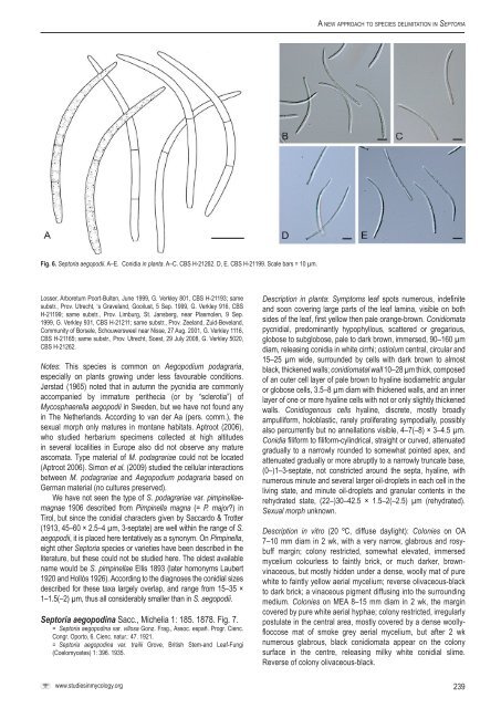
Verkley et al.near the base, holoblastic, proliferat<strong>in</strong>g percurrently 1–many timeswith <strong>in</strong>dist<strong>in</strong>ct annellations, also sympodially, mostly after one ormore percurrent proliferations, 7– 14(–22) × 3–5 µm. Conidia onOA hyal<strong>in</strong>e, pale salmon <strong>in</strong> mass, cyl<strong>in</strong>drical and regularly curved,or abruptly bent <strong>in</strong> the lower or upper cell, gradually attenuated<strong>to</strong> the rounded apex, more abruptly attenuated <strong>in</strong><strong>to</strong> a truncatebase, contents granular with large vacuoles, 1(–3)-septate, not or<strong>in</strong>dist<strong>in</strong>ctly constricted around the septa, contents rich <strong>in</strong> m<strong>in</strong>uteguttulae and granular material, 25.5–41 × (2.0–)2.5–3.0(–4.5) µm.Hosts: On dead leaves and stems of Spergula spp.Material exam<strong>in</strong>ed: Belgium, Beverloo, on dry leaves and stems of Spergulaarvensis, M. Torqu<strong>in</strong>et s.n., isotype BR-MYCO 159328-54, also distributed<strong>in</strong> Westendorp & Wallay, Herb.crypt.Belg., Fasc. 23-24: no.1155. Germany,Brandenburg, Kreis Nieder-Barnim, near Prenden, on leaves and stems of Spergulavernalis, 24 July 1920, H. & P. Sydow s.n., distributed <strong>in</strong> Sydow, Mycothecagermanica 1688, <strong>CBS</strong> H-4765. Netherlands, on Dianthus caryophyllus, Schoutens.n., <strong>CBS</strong> 397.52 (sub S. dianthi Desm.); Prov. Gelderland, ‘t Harde, DoornspijkseHeide, De Zanden, on decay<strong>in</strong>g leaves of Spergula morisonii, A. Aptroot 48300, 13June 2000, epitype designated here <strong>CBS</strong> H-21150 “MBT175350”, liv<strong>in</strong>g cultureex-epitype <strong>CBS</strong> 109010.Notes: This fungus was orig<strong>in</strong>ally described from dry leavesand stems of Spergula arvensis by Westendorp, who describedthe conidia as 30 × 2.5 µm. The type from BR is well-preservedand rich <strong>in</strong> fruitbodies on leaves and stems, where conidia are1(–2)-septate, 20–38 × 2–2.5(–3) µm. The collection Aptroot48300 from Spergula morisonii agrees <strong>in</strong> morphology and can beidentified as conspecific, although it conta<strong>in</strong>s a larger proportionof 2-septate conidia (that are mostly 30–40 µm long) than <strong>in</strong> thetype. The material on Spergula vernalis that was distributed asMycotheca germanica 1688 morphologically also agrees with thesecollections.Other names were later <strong>in</strong>troduced for Sep<strong>to</strong>ria on members ofthe plant genus Spergularia (= Als<strong>in</strong>e), which is closely related <strong>to</strong>Spergula: S. als<strong>in</strong>es Rostr. 1903 from Spergularia sp., conidia 20–31 × 2–3 µm formed <strong>in</strong> 55–120 µm wide pycnidia (Teterevnikova-Babayan 1987; conidia 20–25 × 2–3 µm and 3-septate, <strong>in</strong> theorig<strong>in</strong>al diagnosis of Rostrup 1903, based on material fromAls<strong>in</strong>e verna non Spergula vernalis), S. spergulariae 1903, onSpergularia rubra (conidia 30–45 × 2.5–3 µm, “multiseptate”), S.vandasii 1906, on Als<strong>in</strong>e glomerata, and S. spergular<strong>in</strong>a 1945,on Spergularia longipes (no conidial measurements available).Some of these names could be synonymous with S. spergulae orperhaps S. als<strong>in</strong>es, but <strong>in</strong> order <strong>to</strong> corroborate this, <strong>new</strong> materialneeds <strong>to</strong> be collected and compared <strong>to</strong> the types. Accord<strong>in</strong>g <strong>to</strong>Teterevnikova-Babayan (1987), S. als<strong>in</strong>es differs from S. spergulae<strong>in</strong> conidial shape <strong>in</strong> that the conidial base is more truncate than<strong>in</strong> S. spergulae, and <strong>in</strong> that it is capable of also kill<strong>in</strong>g M<strong>in</strong>uartiaglomerata. Rhabdospora als<strong>in</strong>es Mont. 1892, which was describedfrom dead stems of Als<strong>in</strong>e tenuifolia, is unlikely <strong>to</strong> be conspecificwith S. spergulae, as its conidia were described as 16–18 × 2 µmand 1-septate.Muthumary (1999) studied type material of S. dianthi 1849 (PC344) and by the draw<strong>in</strong>gs he made of it the conidia of this fungusand those of S. spergulae appear very similar <strong>in</strong> shape. Muthumaryreported that the conidia of S. dianthi were 32–48 (av. 40) × 3–4 (av.3) µm, and mostly 1-, rarely 2-septate. Given these measurements,on average, the conidia <strong>in</strong> the type of S. dianthi are clearly longerthan <strong>in</strong> S. spergulae (on average below or around 30). Moreover,S. dianthi is a fungus caus<strong>in</strong>g leaf spots on several Dianthus spp.,while S. spergulae is only known from dry and dead host tissues,and is therefore believed <strong>to</strong> be saprobic (and possibly endophytic).<strong>CBS</strong> 109010 and the only stra<strong>in</strong> available for S. dianthi (<strong>CBS</strong>397.52) show 100 % sequence homology of the LSU, ITS, Btuband Cal, while there are only m<strong>in</strong>or differences <strong>in</strong> Act (99.25 %),EF (97.54 %), and RPB2 (99.42 %). Further work is required <strong>to</strong>establish that S. dianthi and S. spergulae are truely dist<strong>in</strong>ct taxa.Sep<strong>to</strong>ria Sacc., Syll. Fung. 3 : 474. 1884. nom. cons.Type <strong>species</strong> : S. cytisi Desm.A generic description is provided by Quaedvlieg et al. (2013, thisvolume).Sep<strong>to</strong>ria aegopodii Desm. ex J. Kickx, Pl. Crypt. Fland. 1:427. 1876 [Annls Sci. Nat., sér. 6, 7: no 616. 1878?]. Fig. 6.= Sep<strong>to</strong>ria podagrariae Lasch, <strong>in</strong> Rabenh., Herb. mycol. I, no 458. 1843.nomen nudum.= Sphaeria podagrariae Roth, Catal. Bot. 1: 230. 1797.≡ Mycosphaerella podagrariae (Roth : Fr.) Petr., Annls mycol. 19 (3/4):203. 1921.= Cryp<strong>to</strong>sporium aegopodii Preuss, L<strong>in</strong>naea 24: 719 (Fungi Hoyersw., no.322). 1853.≡ Phloeospora aegopodii (Preuss) Grove, British Stem- and Leaf-fungi(Coelomycetes) 1: 434. 1935.≡ Sep<strong>to</strong>ria aegopodii (Preuss) Sacc., Syll. Fung. 3: 529. 1884 [non Desm.1878].?= Sep<strong>to</strong>ria podagrariae var. pimp<strong>in</strong>ellae-magnae Kabát & Bubák, <strong>in</strong> Bubák &Kabát, Ber. naturw.-med. Ver. Innsbruck 30: 19-36 (extr. 11). 1906.= Mycosphaerella aegopodii Potebnia, Annls mycol. 8(1): 49. 1910.Description <strong>in</strong> planta: Symp<strong>to</strong>ms leaf spots numerous but small,angular and delimited by ve<strong>in</strong>lets, visible on both sides of the leaf,white <strong>to</strong> pale yellow. Conidiomata pycnidial, develop<strong>in</strong>g soon afterfirst discolouration of the host tissue, predom<strong>in</strong>antly epiphyllous,mostly also visible from the underside of the lesion, severalscattered <strong>in</strong> each leaf spot, globose <strong>to</strong> subglobose, pale <strong>to</strong> darkbrown (dry<strong>in</strong>g black), immersed, 125–190 µm diam, releas<strong>in</strong>gconidia <strong>in</strong> white cirrhi; ostiolum central, <strong>in</strong>itially circular and 17–35µm wide, later becom<strong>in</strong>g more irregular and up <strong>to</strong> 100 µm wide,surround<strong>in</strong>g cells dark brown, with thickened cell walls; conidiomatalwall except for the part surround<strong>in</strong>g the ostiolum poorly developed,about 10–20 µm thick, composed of pale brown <strong>to</strong> hyal<strong>in</strong>eangular cells 3.5–8 µm diam with th<strong>in</strong> walls. Conidiogenous cellshyal<strong>in</strong>e, discrete, cyl<strong>in</strong>drical <strong>to</strong> narrowly or broadly ampulliform,holoblastic, proliferat<strong>in</strong>g sympodially, 8–15(–18) × 2.5–4.5 µm.Conidia filiform-cyl<strong>in</strong>drical, straight, curved <strong>to</strong> somewhat flexuous,attenuated gradually <strong>to</strong> a relatively broadly rounded apex andbroadly truncate base often provided with a collar of gelat<strong>in</strong>ousmaterial, (0–)1–2(–3)-septate (second and later septa very th<strong>in</strong>and easily overlooked), not constricted around the septa, hyal<strong>in</strong>e,contents with numerous m<strong>in</strong>ute oil-droplets and granular material<strong>in</strong> each cell <strong>in</strong> the liv<strong>in</strong>g state, with m<strong>in</strong>ute oil-droplets and granularcontents <strong>in</strong> the rehydrated state, (30–)55–95(–115) × 3.5–4 µm(liv<strong>in</strong>g; 30–72(–80) × 2.5–4 µm, rehydrated).Description <strong>in</strong> vitro: All attempts <strong>to</strong> grow the isolates from conidiafailed. Some conidia germ<strong>in</strong>ated at the apical cells, but myceliadied with<strong>in</strong> 1–2 d after germ<strong>in</strong>ation.Hosts: Aegopodium podagraria and Pimp<strong>in</strong>ella sp.Material exam<strong>in</strong>ed: Austria, Tirol, Ötztal, Ötz near Habichen, on liv<strong>in</strong>g leaves ofPimp<strong>in</strong>ella sp., 24 July 2000, G. Verkley 1001, <strong>CBS</strong> H-21187. Netherlands, Prov.Overijssel, Losser, <strong>in</strong> garden at Mollenbergstraat, on liv<strong>in</strong>g leaves of Aegopodiumpodagraria, June 1999, G. Verkley 800, <strong>CBS</strong> H-21192; same substr., Prov. Overijssel,238
A <strong>new</strong> <strong>approach</strong> <strong>to</strong> <strong>species</strong> <strong>delimitation</strong> <strong>in</strong> Sep<strong>to</strong>riaFig. 6. Sep<strong>to</strong>ria aegopodii. A–E. Conidia <strong>in</strong> planta. A–C. <strong>CBS</strong> H-21262. D, E. <strong>CBS</strong> H-21199. Scale bars = 10 µm.Losser, Arboretum Poort-Bulten, June 1999, G. Verkley 801, <strong>CBS</strong> H-21193; samesubstr., Prov. Utrecht, ‘s Graveland, Gooilust, 5 Sep. 1999, G. Verkley 916, <strong>CBS</strong>H-21199; same substr., Prov. Limburg, St. Jansberg, near Plasmolen, 9 Sep.1999, G. Verkley 931, <strong>CBS</strong> H-21211; same substr., Prov. Zeeland, Zuid-Beveland,Community of Borsele, Schouwersweel near Nisse, 27 Aug. 2001, G. Verkley 1116,<strong>CBS</strong> H-21165; same substr., Prov. Utrecht, Soest, 29 July 2008, G. Verkley 5020,<strong>CBS</strong> H-21262.Notes: This <strong>species</strong> is common on Aegopodium podagraria,especially on plants grow<strong>in</strong>g under less favourable conditions.Jørstad (1965) noted that <strong>in</strong> autumn the pycnidia are commonlyaccompanied by immature perithecia (or by “sclerotia”) ofMycosphaerella aegopodii <strong>in</strong> Sweden, but we have not found any<strong>in</strong> The Netherlands. Accord<strong>in</strong>g <strong>to</strong> van der Aa (pers. comm.), thesexual morph only matures <strong>in</strong> montane habitats. Aptroot (2006),who studied herbarium specimens collected at high altitudes<strong>in</strong> several localities <strong>in</strong> Europe also did not observe any matureascomata. Type material of M. podagrariae could not be located(Aptroot 2006). Simon et al. (2009) studied the cellular <strong>in</strong>teractionsbetween M. podagrariae and Aegopodium podagraria based onGerman material (no cultures preserved).We have not seen the type of S. podagrariae var. pimp<strong>in</strong>ellaemagnae1906 described from Pimp<strong>in</strong>ella magna (= P. major?) <strong>in</strong>Tirol, but s<strong>in</strong>ce the conidial characters given by Saccardo & Trotter(1913, 45–60 × 2.5–4 µm, 3-septate) are well with<strong>in</strong> the range of S.aegopodii, it is placed here tentatively as a synonym. On Pimp<strong>in</strong>ella,eight other Sep<strong>to</strong>ria <strong>species</strong> or varieties have been described <strong>in</strong> theliterature, but these could not be studied here. The oldest availablename would be S. pimp<strong>in</strong>ellae Ellis 1893 (later homonyms Laubert1920 and Hollós 1926). Accord<strong>in</strong>g <strong>to</strong> the diagnoses the conidial sizesdescribed for these taxa largely overlap, and range from 15–35 ×1–1.5(–2) µm, thus all considerably smaller than <strong>in</strong> S. aegopodii.Sep<strong>to</strong>ria aegopod<strong>in</strong>a Sacc., Michelia 1: 185. 1878. Fig. 7.= Sep<strong>to</strong>ria aegopod<strong>in</strong>a var. villosa Gonz. Frag., Assoc. españ. Progr. Cienc.Congr. Opor<strong>to</strong>, 6. Cienc. natur.: 47. 1921.= Sep<strong>to</strong>ria aegopod<strong>in</strong>a var. trailii Grove, British Stem-and Leaf-Fungi(Coelomycetes) 1: 396. 1935.Description <strong>in</strong> planta: Symp<strong>to</strong>ms leaf spots numerous, <strong>in</strong>def<strong>in</strong>iteand soon cover<strong>in</strong>g large parts of the leaf lam<strong>in</strong>a, visible on bothsides of the leaf, first yellow then pale orange-brown. Conidiomatapycnidial, predom<strong>in</strong>antly hypophyllous, scattered or gregarious,globose <strong>to</strong> subglobose, pale <strong>to</strong> dark brown, immersed, 90–160 µmdiam, releas<strong>in</strong>g conidia <strong>in</strong> white cirrhi; ostiolum central, circular and15–25 µm wide, surrounded by cells with dark brown <strong>to</strong> almostblack, thickened walls; conidiomatal wall 10–28 µm thick, composedof an outer cell layer of pale brown <strong>to</strong> hyal<strong>in</strong>e isodiametric angularor globose cells, 3.5–8 µm diam with thickened walls, and an <strong>in</strong>nerlayer of one or more hyal<strong>in</strong>e cells with not or only slightly thickenedwalls. Conidiogenous cells hyal<strong>in</strong>e, discrete, mostly broadlyampulliform, holoblastic, rarely proliferat<strong>in</strong>g sympodially, possiblyalso percurrently but no annellations visible, 4–7(–8) × 3–4.5 µm.Conidia filiform <strong>to</strong> filiform-cyl<strong>in</strong>drical, straight or curved, attenuatedgradually <strong>to</strong> a narrowly rounded <strong>to</strong> somewhat po<strong>in</strong>ted apex, andattenuated gradually or more abruptly <strong>to</strong> a narrowly truncate base,(0–)1–3-septate, not constricted around the septa, hyal<strong>in</strong>e, withnumerous m<strong>in</strong>ute and several larger oil-droplets <strong>in</strong> each cell <strong>in</strong> theliv<strong>in</strong>g state, and m<strong>in</strong>ute oil-droplets and granular contents <strong>in</strong> therehydrated state, (22–)30–42.5 × 1.5–2(–2.5) µm (rehydrated).Sexual morph unknown.Description <strong>in</strong> vitro (20 ºC, diffuse daylight): Colonies on OA7–10 mm diam <strong>in</strong> 2 wk, with a very narrow, glabrous and rosybuffmarg<strong>in</strong>; colony restricted, somewhat elevated, immersedmycelium colourless <strong>to</strong> fa<strong>in</strong>tly brick, or much darker, brownv<strong>in</strong>aceous,but mostly hidden under a dense, woolly mat of purewhite <strong>to</strong> fa<strong>in</strong>tly yellow aerial mycelium; reverse olivaceous-black<strong>to</strong> dark brick; a v<strong>in</strong>aceous pigment diffus<strong>in</strong>g <strong>in</strong><strong>to</strong> the surround<strong>in</strong>gmedium. Colonies on MEA 8–15 mm diam <strong>in</strong> 2 wk, the marg<strong>in</strong>covered by pure white aerial hyphae; colony restricted, irregularlypostulate <strong>in</strong> the central area, mostly covered by a dense woollyfloccosemat of smoke grey aerial mycelium, but after 2 wknumerous glabrous, black conidiomata appear on the colonysurface <strong>in</strong> the centre, releas<strong>in</strong>g milky white conidial slime.Reverse of colony olivaceous-black.www.studies<strong>in</strong>mycology.org239