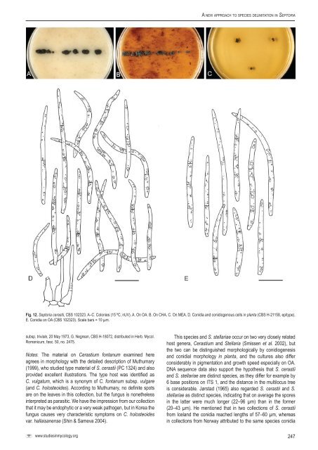
Verkley et al.almost black, glabrous, with a s<strong>in</strong>gle or up <strong>to</strong> 5 ostioli placed on shortpapillae or more elongated necks, that release pale whitish conidialslime; aerial mycelium scanty, diffuse, woolly-floccose, white; reverse<strong>in</strong> the centre most dark slate blue, first surrounded and <strong>in</strong>termixedwith ochreous <strong>to</strong> rust, later more coral. Colonies on CMA 5–9 mmdiam <strong>in</strong> 10 d (24–28 mm <strong>in</strong> 3 wk; > 70 mm <strong>in</strong> 7 wk), with an even,glabrous marg<strong>in</strong>; as on OA but immersed mycelium with a greenishhaze throughout, later almost entirely olivaceous-black; aerialmycelium even more scanty, but higher and reverse darker, darkslate blue throughout most of the colony; conidiomata similar as onOA, but necks shorter or absent. Colonies on MEA 7–9 mm diam <strong>in</strong> 2wk (24–30 mm <strong>in</strong> 3 wk; > 70 mm <strong>in</strong> 7 wk), with an even, undulat<strong>in</strong>g <strong>to</strong>ruffled, glabrous, buff <strong>to</strong> honey marg<strong>in</strong>; colonies first more restricted,pustulate <strong>to</strong> almost conical, but later grow<strong>in</strong>g faster with a planesubmarg<strong>in</strong>al area; immersed mycelium rather dark, near the marg<strong>in</strong>covered by woolly <strong>to</strong> felty white aerial mycelium; mostly composedof spherical conidiomatal <strong>in</strong>itials, superficial mature conidiomatareleas<strong>in</strong>g milky white conidial slime; reverse first dark brick <strong>in</strong> thecentre, near the marg<strong>in</strong> locally grey-olivaceous or c<strong>in</strong>namon, latersepia <strong>to</strong> brown-v<strong>in</strong>aceous, the marg<strong>in</strong> honey. Colonies on CHA 4–10mm diam <strong>in</strong> 10 d (17–32 mm <strong>in</strong> 3 wk; 45–65 mm <strong>in</strong> 7 wk), with anirregular or even, buff marg<strong>in</strong> covered by a diffuse, felty white, latergrey aerial mycelium; further as on MEA, but the colony surfaceless elevated and especially near the marg<strong>in</strong> with greyish, felty <strong>to</strong>tufty aerial mycelium; <strong>in</strong> the centre numerous conidiomata developat the surface, after 3 wk releas<strong>in</strong>g milky white <strong>to</strong> rosy-buff dropletsof conidial slime; reverse <strong>in</strong> the centre blood colour, dark brick <strong>to</strong>c<strong>in</strong>namon at the marg<strong>in</strong>.Conidiogenous cells as <strong>in</strong> planta, but often with relativelylonger necks due <strong>to</strong> repetitive percurrent proliferation. Conidia as <strong>in</strong>planta, but more often 2 and also 3-septate, and mostly 18–34.5 ×1.5–2 µm (OA), 13–32 × 1.5–2 µm (CHA).Hosts: Campanula glomerata, C. takesimana.Material exam<strong>in</strong>ed: Austria, Tirol, Ötztal, Sulztal, Gries, along the river <strong>in</strong> thevillage, on liv<strong>in</strong>g leaves of Campanula glomerata, 1 Aug. 2000, G. Verkley 1034,<strong>CBS</strong> H-21178, liv<strong>in</strong>g cultures <strong>CBS</strong> 109114, 109115. Korea, Taean, on liv<strong>in</strong>g leavesof C. takesimana, H.D. Sh<strong>in</strong>, liv<strong>in</strong>g culture SMKC 21949 = KACC 42622 = <strong>CBS</strong>128589; Daejeon, same substr., H.D. Sh<strong>in</strong>, liv<strong>in</strong>g culture SMKC 24476 = KACC44787 = <strong>CBS</strong> 128604.Notes: The first <strong>species</strong> described on Campanula is S. campanulae,for which Sh<strong>in</strong> & Sameva (2004) provided a detailed descriptionbased on material occurr<strong>in</strong>g <strong>in</strong> Korea on C. punctata and C.takeshimana (conidia mostly 1-septate, 13–24 × 1.5–2 µm). Sh<strong>in</strong>& Sameva summerised the his<strong>to</strong>ry of the Sep<strong>to</strong>ria <strong>species</strong> on thegenus Campanula. Of the three <strong>species</strong> most often accepted, viz.,S. campanulae, S. obscura, and S. trachelii, S. campanulae fits thecurrent material best. Sep<strong>to</strong>ria arcautei was not mentioned by Sh<strong>in</strong>& Sameva. This <strong>species</strong> was described from C. glomerata <strong>in</strong> Spa<strong>in</strong>,and accord<strong>in</strong>g <strong>to</strong> the orig<strong>in</strong>al description by Unanumo, the pycnidiaare predom<strong>in</strong>antly epiphyllous, 55.8–74.8 µm diam, and the conidiacont<strong>in</strong>uous, 20–25.7 × 0.8 µm. Sep<strong>to</strong>ria campanulae is closely related<strong>to</strong> several <strong>species</strong> from hosts <strong>in</strong> Apiaceae, <strong>in</strong>clud<strong>in</strong>g S. aegopod<strong>in</strong>a,S. oenanthis, and S. sii (Fig. 2). Sequenc<strong>in</strong>g results of <strong>CBS</strong> 109114and 109115 were puzzl<strong>in</strong>g, suggest<strong>in</strong>g possible contam<strong>in</strong>ation.Sep<strong>to</strong>ria cerastii Roberge ex Desm., Annls Sci. Nat., sér. 3,Bot. 11: 347. 1849. Fig. 12.Description <strong>in</strong> planta: Symp<strong>to</strong>ms <strong>in</strong>def<strong>in</strong>ite, yellow <strong>to</strong> brown leafspots, but more often on wither<strong>in</strong>g parts of leaves, stems and bracts.Conidiomata pycnidial, on leaves amphigenous but predom<strong>in</strong>atelyepiphyllous, scattered or aggregated, globose, semi-immersed,80–125(–150) µm diam; ostiolum circular, central, 20–45 µm wide,surround<strong>in</strong>g cells somewhat darker; conidiomatal wall composedof textura angularis without dist<strong>in</strong>ctly differentiated layers, theouter cells with brown, somewhat thickened walls and 4–6.5 µmdiam, the <strong>in</strong>ner cells hyal<strong>in</strong>e and th<strong>in</strong>-walled and 3.5–6 µm diam.Conidiogenous cells ampulliform, or elongated ampulliform with adist<strong>in</strong>ct neck, hyal<strong>in</strong>e, holoblastic, proliferat<strong>in</strong>g percurrently 1–manytimes with <strong>in</strong>dist<strong>in</strong>ct annellations, also sympodially, 5–10 × 3–5 µm.Conidia filiform <strong>to</strong> filiform-cyl<strong>in</strong>drical, straight, curved, or flexuous,gradually attenuated <strong>to</strong> a rounded or more or less po<strong>in</strong>ted apex,abruptly attenuated <strong>in</strong><strong>to</strong> a truncate base, (1–)2–4(–5)-septate,not or <strong>in</strong>dist<strong>in</strong>ctly constricted around the septa, hyal<strong>in</strong>e, contentsmoderately rich <strong>in</strong> small guttulae, m<strong>in</strong>utely granular material andlarge vacuoles <strong>in</strong> the liv<strong>in</strong>g state, <strong>in</strong> the rehydrated state with<strong>in</strong>conspicuous contents and no oil-droplets, (21–)30–52(–57) ×1.5–2 µm (rehydrated). Sexual morph unknown.Description <strong>in</strong> vitro: Colonies on OA 2–4 mm diam <strong>in</strong> 2 wk (10–13mm <strong>in</strong> 6 wk), the marg<strong>in</strong> irregular <strong>to</strong> ruffled, almost as dark as res<strong>to</strong>f the colony, covered by diffuse, grey aerial mycelium; the colonyspread<strong>in</strong>g, almost plane <strong>to</strong> somewhat irregularly lifted and pustulate,immersed mycelium olivaceous-black <strong>to</strong> black, covered with dense,grey, woolly aerial mycelium; conidiomata start<strong>in</strong>g <strong>to</strong> develop at thesurface after 10–15 d; reverse olivaceous-black. Colonies on CMA2–5 mm diam <strong>in</strong> 2 wk (13–17 mm <strong>in</strong> 6 wk), as on OA; conidialslime milky white; reverse greenish grey <strong>to</strong> almost black. Colonieson MEA 0.5–1.5 mm diam <strong>in</strong> 2 wk (4–6 mm <strong>in</strong> 6 wk), as on OA,with equally dense and long, woolly, grey aerial mycelium; colonyhemispherical, with scarce pycnidial conidiomata develop<strong>in</strong>gtardily; reverse dark slate blue <strong>to</strong> black. Colonies on CHA 1–3 mmdiam <strong>in</strong> 2 wk (8–12 mm <strong>in</strong> 6 wk), as on OA, but colonies moredist<strong>in</strong>ctly lifted above the agar surface, hemispherical, and aerialmycelium denser but shorter; conidiomata develop<strong>in</strong>g scarcely atthe surface.Conidiomata pycnidial and similar as <strong>in</strong> planta, 100–150 µmdiam, or merged <strong>in</strong><strong>to</strong> larger complexes especially on the agarsurface, dark olivaceous-black <strong>to</strong> black, up <strong>to</strong> 250 µm diam; ostiolumas <strong>in</strong> planta, or absent; Conidiogenous cells hyal<strong>in</strong>e, ampuliform, orelongated ampulliform <strong>to</strong> cyl<strong>in</strong>drical, with a dist<strong>in</strong>ct neck, holoblastic,proliferat<strong>in</strong>g percurrently 1–many times with <strong>in</strong>dist<strong>in</strong>ct scars(annellations), also sympodially, 5–12(–15) × 3–5(–6.5) µm. Conidiaon OA similar as <strong>in</strong> planta, 1–3(–5)-septate, <strong>in</strong>dist<strong>in</strong>ctly constrictedaround the septa, hyal<strong>in</strong>e, contents moderately rich <strong>in</strong> small guttulae,m<strong>in</strong>utely granular material and large vacuoles <strong>in</strong> the liv<strong>in</strong>g state,(26–)35–50(–57) × 1.5–2.5 µm (T), released from superficialconidiomata <strong>in</strong> whitish cirrhi or slimy masses.Hosts: In leaf spots and on wither<strong>in</strong>g leaves, stems and bracts ofCerastium spp. Accord<strong>in</strong>g <strong>to</strong> Markevičius & Treigienė (2003), alsoon Stellaria holostea.Material exam<strong>in</strong>ed: Korea, Hoengseong, on C. holosteoides var. hallaisanense,14 May 2006, H.D. Sh<strong>in</strong>, <strong>CBS</strong> 128586 = KACC 42367 = SMKC 21781; sameloc., substr., H.D. Sh<strong>in</strong>, <strong>CBS</strong> 128612 = KACC 42831 = SMKC 22609; Jeju, onC. holosteoides, 1 Nov. 2007, H.D. Sh<strong>in</strong>, <strong>CBS</strong> 128626 = KACC 43220 = SMKC23137. Netherlands, prov. Utrecht, Baarn, on liv<strong>in</strong>g leaves of Cerastium sp., 9 Aug.1968, H.A. van der Aa 731, <strong>CBS</strong> H-18069; same loc., substratum, 18 Oct. 1962,H.A. van der Aa, <strong>CBS</strong> H-18070, and 19 Oct. 1963, <strong>CBS</strong> H-18071; Prov. NoordHolland, Amsterdamse Waterleid<strong>in</strong>gdu<strong>in</strong>en, near Ruigeveld, on wither<strong>in</strong>g leavesof Cerastium fontanum subsp. vulgare, 31 Aug. 1999, G. Verkley & A. van Iperen915, epitype designated here <strong>CBS</strong> H-21158 “MBT175351”, liv<strong>in</strong>g culture ex-epitype<strong>CBS</strong> 102323. Romania, distr. Ilfov, Malu-Spart, on liv<strong>in</strong>g leaves of C. fontanum246
A <strong>new</strong> <strong>approach</strong> <strong>to</strong> <strong>species</strong> <strong>delimitation</strong> <strong>in</strong> Sep<strong>to</strong>riaFig. 12. Sep<strong>to</strong>ria cerastii, <strong>CBS</strong> 102323. A–C. Colonies (15 ºC, nUV). A. On OA. B. On CHA. C. On MEA. D. Conidia and conidiogenous cells <strong>in</strong> planta (<strong>CBS</strong> H-21158, epitype).E. Conidia on OA (<strong>CBS</strong> 102323). Scale bars = 10 µm.subsp. triviale, 20 May 1973, G. Negrean, <strong>CBS</strong> H-18072, distributed <strong>in</strong> Herb. Mycol.Romanicum, fasc. 50, no. 2475.Notes: The material on Cerastium fontanum exam<strong>in</strong>ed hereagrees <strong>in</strong> morphology with the detailed description of Muthumary(1999), who studied type material of S. cerastii (PC 1324) and alsoprovided excellent illustrations. The type host was identified asC. vulgatum, which is a synonym of C. fontanum subsp. vulgare(and C. holosteoides). Accord<strong>in</strong>g <strong>to</strong> Muthumary, no def<strong>in</strong>ite spotsare on the leaves <strong>in</strong> this collection, but the fungus is nonetheless<strong>in</strong>terpreted as parasitic. We have the impression from our collectionthat it may be endophytic or a very weak pathogen, but <strong>in</strong> Korea thefungus causes very characteristic symp<strong>to</strong>ms on C. holosteoidesvar. hallaisanense (Sh<strong>in</strong> & Sameva 2004).This <strong>species</strong> and S. stellariae occur on two very closely relatedhost genera, Cerastium and Stellaria (Smissen et al. 2002), butthe two can be dist<strong>in</strong>guished morphologically by conidiogenesisand conidial morphology <strong>in</strong> planta, and the cultures also differconsiderably <strong>in</strong> pigmentation and growth speed especially on OA.DNA sequence data also support the hypothesis that S. cerastiiand S. stellariae are dist<strong>in</strong>ct <strong>species</strong>, as they differ for example by6 base positions on ITS 1, and the distance <strong>in</strong> the multilocus treeis considerable. Jørstad (1965) also regarded S. cerastii and S.stellariae as dist<strong>in</strong>ct <strong>species</strong>, <strong>in</strong>dicat<strong>in</strong>g that on average the spores<strong>in</strong> the latter were much longer (22–96 µm) than <strong>in</strong> the former(20–43 µm). He mentioned that <strong>in</strong> two collections of S. cerastiifrom Iceland the conidia reached lengths of 57–60 µm, whereas<strong>in</strong> collections from Norway attributed <strong>to</strong> the same <strong>species</strong> conidiawww.studies<strong>in</strong>mycology.org247