

A new approach to species delimitation in Septoria - CBS - KNAW
A new approach to species delimitation in Septoria - CBS - KNAW
A new approach to species delimitation in Septoria - CBS - KNAW

Create successful ePaper yourself
Turn your PDF publications into a flip-book with our unique Google optimized e-Paper software.
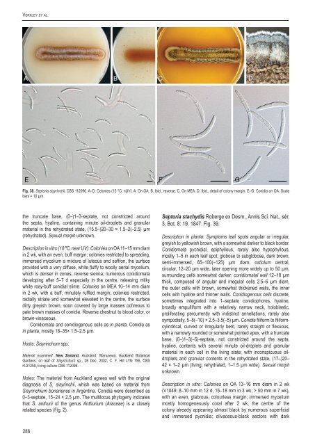
Verkley et al.Fig. 38. Sep<strong>to</strong>ria sisyr<strong>in</strong>chii, <strong>CBS</strong> 112096. A–D. Colonies (15 °C, nUV). A. On OA. B. Ibid., reverse. C. On MEA. D. Ibid., detail of colony marg<strong>in</strong>. E–G. Conidia on OA. Scalebars = 10 µm.the truncate base, (0–)1–3-septate, not constricted aroundthe septa, hyal<strong>in</strong>e, conta<strong>in</strong><strong>in</strong>g m<strong>in</strong>ute oil-droplets and granularmaterial <strong>in</strong> the rehydrated state, (15.5–)20–30 × 1.5–2(–2.5) µm(rehydrated). Sexual morph unknown.Description <strong>in</strong> vitro (18 ºC, near UV): Colonies on OA 11–15 mm diam<strong>in</strong> 2 wk, with an even, buff marg<strong>in</strong>; colonies restricted <strong>to</strong> spread<strong>in</strong>g,immersed mycelium a mixture of luteous and saffron, the surfaceprovided with a very diffuse, white fluffy <strong>to</strong> woolly aerial mycelium,which is denser <strong>in</strong> zones; reverse sienna; numerous conidiomatadevelop<strong>in</strong>g after 5–7 d especially <strong>in</strong> the centre, releas<strong>in</strong>g milkywhite rosy-buff conidial slime. Colonies on MEA 10–14 mm diam<strong>in</strong> 2 wk, with a buff, m<strong>in</strong>utely ruffled marg<strong>in</strong>; colonies restricted,radially striate and somewhat elevated <strong>in</strong> the centre, the surfacedirty greyish brown, soon covered by large masses ochreous <strong>to</strong>pale brown masses of conidia. Reverse chestnut <strong>to</strong> blood color, orbrown-v<strong>in</strong>aceous.Conidiomata and conidiogenous cells as <strong>in</strong> planta. Conidia as<strong>in</strong> planta, mostly 18–35× 1.5–2.5 µm.Hosts: Sisyr<strong>in</strong>chum spp.Material exam<strong>in</strong>ed: New Zealand, Auckland, Manurewa, Auckland BotanicalGardens, on leaf of Sisyr<strong>in</strong>chum sp., 28 Dec. 2002, C. F. Hill LYN 755, <strong>CBS</strong>H-21259, liv<strong>in</strong>g culture <strong>CBS</strong> 112096.Notes: The material from Auckland agrees well with the orig<strong>in</strong>aldiagnosis of S. sisyr<strong>in</strong>chii, which was based on material fromSisyr<strong>in</strong>chium bonariense <strong>in</strong> Argent<strong>in</strong>a. Conidia were described as0–3-septate, 15–24 × 2.5 µm. The multilocus phylogeny <strong>in</strong>dicatesthat S. anthurii of the genus Anthurium (Araceae) is a closelyrelated <strong>species</strong> (Fig. 2).Sep<strong>to</strong>ria stachydis Roberge ex Desm., Annls Sci. Nat., sér.3, Bot. 8: 19. 1847. Fig. 39.Description <strong>in</strong> planta: Symp<strong>to</strong>ms leaf spots angular or irregular,greyish <strong>to</strong> yellowish brown, with a somewhat darker <strong>to</strong> black border.Conidiomata pycnidial, epiphyllous, rarely also hypophyllous,mostly 1–5 <strong>in</strong> each leaf spot, globose <strong>to</strong> subglobose, dark brown,semi-immersed, 65–100(–125) µm diam; ostiolum central,circular, 12–20 µm wide, later open<strong>in</strong>g more widely up <strong>to</strong> 50 µm,surround<strong>in</strong>g cells somewhat darker; conidiomatal wall 12–18 µmthick, composed of angular and irregular cells 2.5–6 µm diam,the outer cells with brown, somewhat thickened walls, the <strong>in</strong>nercells with hyal<strong>in</strong>e and th<strong>in</strong>ner walls. Conidiogenous cells discrete,sometimes <strong>in</strong>tegrated <strong>in</strong><strong>to</strong> 1–septate conidiophores, hyal<strong>in</strong>e,broadly ampulliform with a relatively narrow neck, holoblastic,proliferat<strong>in</strong>g percurrently with <strong>in</strong>dist<strong>in</strong>ct annellations, rarely alsosympodially, 5–8(–10) × 2.5–3.5(–5) µm. Conidia filiform <strong>to</strong> filiformcyl<strong>in</strong>drical,curved or irregularly bent, rarely straight or flexuous,with a narrowly rounded or somewhat po<strong>in</strong>ted apex, with a truncatebase, (0–)1–3(–5)-septate, not constricted around the septa,hyal<strong>in</strong>e, contents with several m<strong>in</strong>ute oil-droplets and granularmaterial <strong>in</strong> each cell <strong>in</strong> the liv<strong>in</strong>g state, with <strong>in</strong>conspicuous oildropletsand granular contents <strong>in</strong> the rehydrated state, (17–)20–42 × 1–2 µm (liv<strong>in</strong>g; rehydrated, 1–1.5 µm wide). Sexual morphunknown.Description <strong>in</strong> vitro: Colonies on OA 13–16 mm diam <strong>in</strong> 2 wk(V1049: 8–10 mm <strong>in</strong> 12 d, 16–18 mm <strong>in</strong> 3 wk; > 50 mm <strong>in</strong> 7 wk),with an even, glabrous, colourless marg<strong>in</strong>; immersed myceliummostly homogeneously coral after 2 wk, the centre of thecolony already appear<strong>in</strong>g almost black by numerous superficialand immersed pycnidia; olivaceous-black sec<strong>to</strong>rs with dark288


