

A new approach to species delimitation in Septoria - CBS - KNAW
A new approach to species delimitation in Septoria - CBS - KNAW
A new approach to species delimitation in Septoria - CBS - KNAW

You also want an ePaper? Increase the reach of your titles
YUMPU automatically turns print PDFs into web optimized ePapers that Google loves.
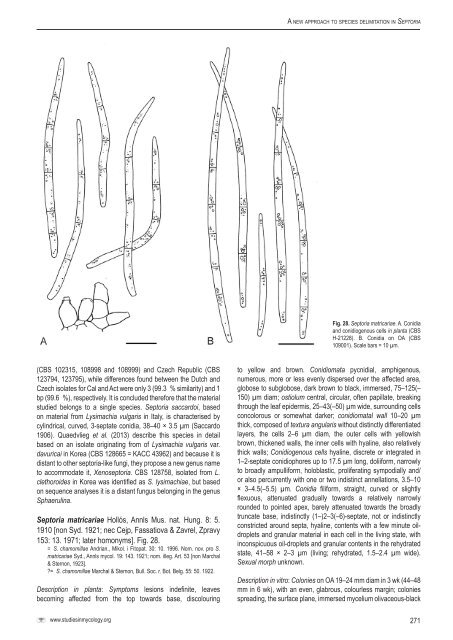
A <strong>new</strong> <strong>approach</strong> <strong>to</strong> <strong>species</strong> <strong>delimitation</strong> <strong>in</strong> Sep<strong>to</strong>riaFig. 28. Sep<strong>to</strong>ria matricariae. A. Conidiaand conidiogenous cells <strong>in</strong> planta (<strong>CBS</strong>H-21228). B. Conidia on OA (<strong>CBS</strong>109001). Scale bars = 10 µm.(<strong>CBS</strong> 102315, 108998 and 108999) and Czech Republic (<strong>CBS</strong>123794, 123795), while differences found between the Dutch andCzech isolates for Cal and Act were only 3 (99.3 % similarity) and 1bp (99.6 %), respectively. It is concluded therefore that the materialstudied belongs <strong>to</strong> a s<strong>in</strong>gle <strong>species</strong>. Sep<strong>to</strong>ria saccardoi, basedon material from Lysimachia vulgaris <strong>in</strong> Italy, is characterised bycyl<strong>in</strong>drical, curved, 3-septate conidia, 38–40 × 3.5 µm (Saccardo1906). Quaedvlieg et al. (2013) describe this <strong>species</strong> <strong>in</strong> detailbased on an isolate orig<strong>in</strong>at<strong>in</strong>g from of Lysimachia vulgaris var.davuricai <strong>in</strong> Korea (<strong>CBS</strong> 128665 = KACC 43962) and because it isdistant <strong>to</strong> other sep<strong>to</strong>ria-like fungi, they propose a <strong>new</strong> genus name<strong>to</strong> accommodate it, Xenosep<strong>to</strong>ria. <strong>CBS</strong> 128758, isolated from L.clethoroides <strong>in</strong> Korea was identified as S. lysimachiae, but basedon sequence analyses it is a distant fungus belong<strong>in</strong>g <strong>in</strong> the genusSphaerul<strong>in</strong>a.Sep<strong>to</strong>ria matricariae Hollós, Annls Mus. nat. Hung. 8: 5.1910 [non Syd. 1921; nec Cejp, Fassatiova & Zavrel, Zpravy153: 13. 1971; later homonyms]. Fig. 28.= S. chamomillae Andrian., Mikol. i Fi<strong>to</strong>pat. 30: 10. 1996. Nom. nov. pro S.matricariae Syd., Annls mycol. 19: 143. 1921; nom. illeg. Art. 53 [non Marchal& Sternon, 1923].?= S. chamomillae Marchal & Sternon, Bull. Soc. r. Bot. Belg. 55: 50. 1922.Description <strong>in</strong> planta: Symp<strong>to</strong>ms lesions <strong>in</strong>def<strong>in</strong>ite, leavesbecom<strong>in</strong>g affected from the <strong>to</strong>p <strong>to</strong>wards base, discolour<strong>in</strong>g<strong>to</strong> yellow and brown. Conidiomata pycnidial, amphigenous,numerous, more or less evenly dispersed over the affected area,globose <strong>to</strong> subglobose, dark brown <strong>to</strong> black, immersed, 75–125(–150) µm diam; ostiolum central, circular, often papillate, break<strong>in</strong>gthrough the leaf epidermis, 25–43(–50) µm wide, surround<strong>in</strong>g cellsconcolorous or somewhat darker; conidiomatal wall 10–20 µmthick, composed of textura angularis without dist<strong>in</strong>ctly differentiatedlayers, the cells 2–6 µm diam, the outer cells with yellowishbrown, thickened walls, the <strong>in</strong>ner cells with hyal<strong>in</strong>e, also relativelythick walls; Conidiogenous cells hyal<strong>in</strong>e, discrete or <strong>in</strong>tegrated <strong>in</strong>1–2-septate conidiophores up <strong>to</strong> 17.5 µm long, doliiform, narrowly<strong>to</strong> broadly ampulliform, holoblastic, proliferat<strong>in</strong>g sympodially and/or also percurrently with one or two <strong>in</strong>dist<strong>in</strong>ct annellations, 3.5–10× 3–4.5(–5.5) µm. Conidia filiform, straight, curved or slightlyflexuous, attenuated gradually <strong>to</strong>wards a relatively narrowlyrounded <strong>to</strong> po<strong>in</strong>ted apex, barely attenuated <strong>to</strong>wards the broadlytruncate base, <strong>in</strong>dist<strong>in</strong>ctly (1–)2–3(–6)-septate, not or <strong>in</strong>dist<strong>in</strong>ctlyconstricted around septa, hyal<strong>in</strong>e, contents with a few m<strong>in</strong>ute oildropletsand granular material <strong>in</strong> each cell <strong>in</strong> the liv<strong>in</strong>g state, with<strong>in</strong>conspicuous oil-droplets and granular contents <strong>in</strong> the rehydratedstate, 41–58 × 2–3 µm (liv<strong>in</strong>g; rehydrated, 1.5–2.4 µm wide).Sexual morph unknown.Description <strong>in</strong> vitro: Colonies on OA 19–24 mm diam <strong>in</strong> 3 wk (44–48mm <strong>in</strong> 6 wk), with an even, glabrous, colourless marg<strong>in</strong>; coloniesspread<strong>in</strong>g, the surface plane, immersed mycelium olivaceous-blackwww.studies<strong>in</strong>mycology.org271


