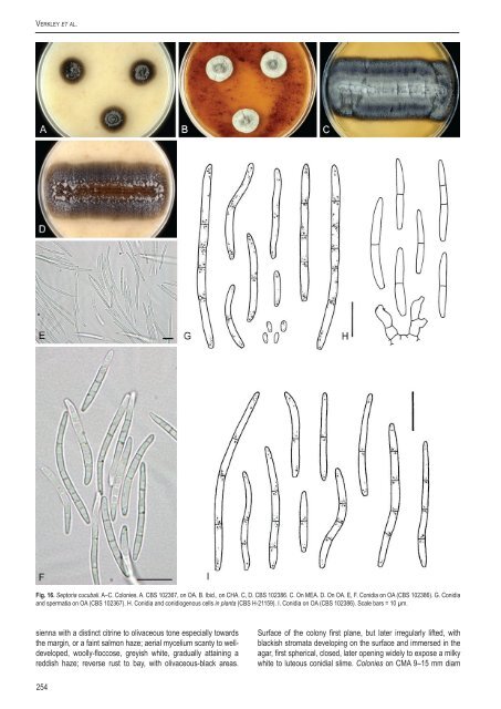
Verkley et al.Fig. 16. Sep<strong>to</strong>ria cucubali. A–C. Colonies. A. <strong>CBS</strong> 102367, on OA. B. Ibid., on CHA. C, D. <strong>CBS</strong> 102386. C. On MEA. D. On OA. E, F. Conidia on OA (<strong>CBS</strong> 102386). G. Conidiaand spermatia on OA (<strong>CBS</strong> 102367). H. Conidia and conidiogenous cells <strong>in</strong> planta (<strong>CBS</strong> H-21159). I. Conidia on OA (<strong>CBS</strong> 102386). Scale bars = 10 µm.sienna with a dist<strong>in</strong>ct citr<strong>in</strong>e <strong>to</strong> olivaceous <strong>to</strong>ne especially <strong>to</strong>wardsthe marg<strong>in</strong>, or a fa<strong>in</strong>t salmon haze; aerial mycelium scanty <strong>to</strong> welldeveloped,woolly-floccose, greyish white, gradually atta<strong>in</strong><strong>in</strong>g areddish haze; reverse rust <strong>to</strong> bay, with olivaceous-black areas.Surface of the colony first plane, but later irregularly lifted, withblackish stromata develop<strong>in</strong>g on the surface and immersed <strong>in</strong> theagar, first spherical, closed, later open<strong>in</strong>g widely <strong>to</strong> expose a milkywhite <strong>to</strong> luteous conidial slime. Colonies on CMA 9–15 mm diam254
A <strong>new</strong> <strong>approach</strong> <strong>to</strong> <strong>species</strong> <strong>delimitation</strong> <strong>in</strong> Sep<strong>to</strong>ria<strong>in</strong> 2 wk (43–45 mm <strong>in</strong> 6 wk), with an even, glabrous, colourless<strong>to</strong> buff marg<strong>in</strong>; further as on OA, but immersed mycelium only <strong>in</strong>the centre sienna, for the most olivaceous <strong>to</strong> almost dull green;aerial mycelium similar <strong>in</strong> colour and texture, but scarcer; reverseolivaceous-black, with dist<strong>in</strong>ct rust central areas; conidiomata lessdeveloped. Colonies on MEA 9–16 mm diam <strong>in</strong> 2 wk, with an even,buff or peach <strong>to</strong> scarlet marg<strong>in</strong>, mostly hidden under tufts of aerialmycelium; colonies hemispherical, sometimes radially striate,immersed mycelium dark ochreous <strong>to</strong> greyish brown or olivaceousblack,mostly covered by f<strong>in</strong>ely felty or floccose-tufty, white, greyishor scarlet aerial mycelium; luteous <strong>to</strong> reddish diffusable pigmentsometimes present; reverse rust <strong>to</strong> chestnut, marg<strong>in</strong> apricot;stromata scarcely develop<strong>in</strong>g, releas<strong>in</strong>g milky white <strong>to</strong> rosy-buffconidial slime. Colonies on CHA (4–)6–9 mm diam <strong>in</strong> 2 wk [(30–)40–46 mm <strong>in</strong> 6 wk], as on MEA, conidial slime first rosy-buff, laterochreous.Conidiomata pycnidial, as <strong>in</strong> planta but often larger, 100–175µm, or merg<strong>in</strong>g <strong>in</strong><strong>to</strong> larger complexes; conidiogenous cells as<strong>in</strong> planta, but annellations more dist<strong>in</strong>ct. Conidia fusiform-cyl<strong>in</strong>drical<strong>to</strong> cyl<strong>in</strong>drical, straight or weakly curved, gradually attenuated <strong>to</strong> arounded or more or less po<strong>in</strong>ted apex, abruptly attenuated <strong>in</strong><strong>to</strong>a narrow, truncate base, (0–)1–3(–4)-septate, not or <strong>in</strong>dist<strong>in</strong>ctlyconstricted around the septa, hyal<strong>in</strong>e, contents m<strong>in</strong>utely granularwith small oil-droplets, (9–)15–29(–52) × 2–2.5 µm.Both on the plant and <strong>in</strong> culture sperma<strong>to</strong>gonia of an Asteromellastate were produced, <strong>in</strong> which 0-septate, ellipsoid spermatia wereformed 2–3 × 1–1.5 µm. No sexual morph was observed.Hosts: on liv<strong>in</strong>g leaves of Cucubalus baccifer and Saponariaoffic<strong>in</strong>alis.Material exam<strong>in</strong>ed: Germany, isolated from leaf litter of Fagus sylvatica, M.Unterseher, liv<strong>in</strong>g culture <strong>CBS</strong> 124874. Netherlands, Prov. Gelderland, Mill<strong>in</strong>genaan de Rijn, Mill<strong>in</strong>gerwaard, on liv<strong>in</strong>g leaves of Cucubalus baccifer, 6 Oct. 1999, G.Verkley 941, <strong>CBS</strong> H-21159, liv<strong>in</strong>g cultures <strong>CBS</strong> 102367, 102368; same loc., date,brown leaf marg<strong>in</strong> on liv<strong>in</strong>g leaves of Saponaria offic<strong>in</strong>alis, 6 Oct. 1999, G. Verkley938, <strong>CBS</strong> H-21218, liv<strong>in</strong>g culture <strong>CBS</strong> 102386.Notes: The material on Cucubalus available for this study showedconidia (9–)15–19(–23) × 2–2.5 µm, thus much shorter andsomewhat narrower than reported for S. cucubali <strong>in</strong> the orig<strong>in</strong>aldiagnosis (34–50 × 1.5–2 µm; based on material collected <strong>in</strong>July), and by Teterevnikova-Babayan (1987). This Dutch materialwas collected much later <strong>in</strong> the season than the type, and underrelatively dry conditions. Averages of conidial width and especiallylengths seen <strong>in</strong> specimens collected under adverse conditionssuch as drought or cold can be lower as compared <strong>to</strong> materialcollected under optimal conditions. The isolates obta<strong>in</strong>ed from thismaterial were, however, capable of produc<strong>in</strong>g conidia up <strong>to</strong> 52 µm<strong>in</strong> length. This would be <strong>in</strong> good agreement with S. cucubali, as arethe morphology of the pycnidia, the shape and width of the conidia,as well as the symp<strong>to</strong>ms on the plant described by Teterevnikova-Babayan (1987) for S. cucubali. Markevičius & Treigiene (2003)reported S. dimera on Cucubalus, and that <strong>species</strong> is characterisedby conidia that are wider (21–35 × 3.2–4.3 µm; Vanev et al. 1997report 26–65 × 2.5–4 µm for that <strong>species</strong>).The isolates from Cucubalus were also very similar <strong>to</strong> thoseobta<strong>in</strong>ed from the material collected <strong>in</strong> the same area on Saponaria,and the sequences obta<strong>in</strong>ed <strong>in</strong>dicate that these isolates all belong<strong>to</strong> a s<strong>in</strong>gle <strong>species</strong>. The material on the plant studied here differsfrom the description of S. saponariae provided by Teterevnikova-Babayan (1987), who describes conidia as 1–3-septate, 25–59 ×3.3–4.5 µm. That <strong>species</strong> thus has much wider conidia. Host rangeof S. cucubali <strong>in</strong> literature only mentions Cucubalus, but it is clearfrom the present study that it also <strong>in</strong>cludes Saponaria offic<strong>in</strong>alis.The stra<strong>in</strong> isolated from beech leaf litter may be an accidentaldweller and orig<strong>in</strong>ate from a Caryophyllaceae host grow<strong>in</strong>g <strong>in</strong> thevic<strong>in</strong>ity. That the fungus would be capable of <strong>in</strong>fect<strong>in</strong>g Fagus leavesas an endophyte seems unlikely but cannot be excluded.Sep<strong>to</strong>ria cucurbitacearum Sacc., Nuovo G. bot. ital. 8: 205.1876.Description <strong>in</strong> vitro: Colonies on OA 38 mm diam <strong>in</strong> 5 wk, with aneven, or slightly undulat<strong>in</strong>g, colourless, glabrous marg<strong>in</strong>; coloniesrestricted <strong>to</strong> moderately spread<strong>in</strong>g, almost entirely olivaceousblack,due <strong>to</strong> brown-walled immersed hyphae, the surface mostlyglabrous, yet <strong>in</strong> the centre and around pycnidia often with greyishwhite, pru<strong>in</strong>ose aerial hyphae. Conidiomata numerous, scattered orgregarious, black, pycnidial, with a s<strong>in</strong>gle often quite long ostiolateneck, but fruitbodies often burst<strong>in</strong>g somewhere <strong>in</strong> the lower wall,conidial slime pale white; reverse concolourous. Conidiogenouscells hyal<strong>in</strong>e, discrete, ampulliform <strong>to</strong> cyl<strong>in</strong>drical, holoblastic, with1–3 percurrent proliferations, 8–16 × 3.5–5 µm. Conidia filiform,curved or flexuous, hyal<strong>in</strong>e, 3–5(–7)-septate, not constrictedaround the septa, narrowly rounded at the <strong>to</strong>p, slighty attenuat<strong>in</strong>g<strong>to</strong> a narrowly truncate base, with m<strong>in</strong>ute oil-droplets, (30–)35–55(–72) × 1.5–2(–2.5) µm.Hosts: Cucurbita spp., Cucumis spp. and Citrullus vulgaris.Material exam<strong>in</strong>ed: New Zealand, culture isolated from liv<strong>in</strong>g leaves of Cucurbitamaxima, date of collection and isolation unknown (deposited <strong>in</strong> Feb. 1977), H. J.Boesew<strong>in</strong>kel s.n., <strong>CBS</strong> 178.77.Notes: No specimens on plant material were available for this study.A description based on specimens from Cucumis, Cucurbita andCitrullus collected <strong>in</strong> Australia is provided by Priest (2006), and thesporulat<strong>in</strong>g structures observed <strong>in</strong> <strong>CBS</strong> 178.77 on OA agree wellwith that description. Sep<strong>to</strong>ria cucurbitacearum is the oldest nameon plants of the family Cucurbitaceae, and Punithal<strong>in</strong>gam (1982)discussed the relationship with the other taxa on the host generaCucurbita and Cucumis. On the basis of the multilocus sequenceanalysis it can be concluded that S. cucurbitacearum is closelyrelated <strong>to</strong> S. lycospersici (<strong>CBS</strong> 354.49 and 128654), S. malagutii(<strong>CBS</strong> 106.80), and S. apiicola.Sep<strong>to</strong>ria digitalis Pass., Atti Soc. crit<strong>to</strong>g. ital. 2: 36. 1879.Fig. 17.Description <strong>in</strong> planta (based on <strong>CBS</strong> H-18090): Symp<strong>to</strong>ms leafspots hologenous, scattered, circular <strong>to</strong> elliptical, pale yellowishbrown, def<strong>in</strong>ite with a dark brown border, or <strong>in</strong>def<strong>in</strong>ite, surroundedby a larger area of the leaf which turns reddish purple. Conidiomatapycnidial, epiphyllous, numerous scattered <strong>in</strong> each leaf spot,subglobose <strong>to</strong> globose, immersed, brown <strong>to</strong> black, (70–)85–130µm diam; ostiolum central, <strong>in</strong>itially circular and 20–45 µm wide,later more irregular and up <strong>to</strong> 60 µm wide, surround<strong>in</strong>g cellsundifferentiated; conidiomatal wall about 12.5–20 µm thick,composed of an outer layer of isodiametric cells 4.5–8(–10) µmdiam or more irregular cells with brown walls 1–2 µm thick, andan <strong>in</strong>ner layer of angular <strong>to</strong> globose cells 2.5–4(–6) µm diam withrelatively th<strong>in</strong>, hyal<strong>in</strong>e walls. Conidiogenous cells hyal<strong>in</strong>e, discrete,rarely <strong>in</strong>tegrated <strong>in</strong> 1-septate conidiophores, globose, doliiform orwww.studies<strong>in</strong>mycology.org255