

A new approach to species delimitation in Septoria - CBS - KNAW
A new approach to species delimitation in Septoria - CBS - KNAW
A new approach to species delimitation in Septoria - CBS - KNAW

You also want an ePaper? Increase the reach of your titles
YUMPU automatically turns print PDFs into web optimized ePapers that Google loves.
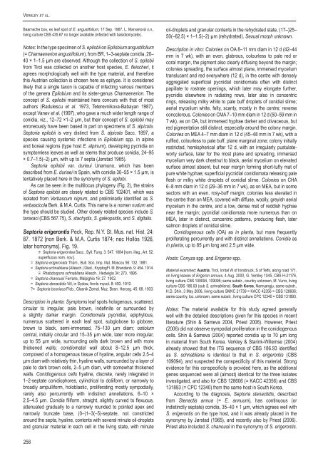
Verkley et al.Baarnsche bos, ex leaf spot of E. angustifolium, 17 Sep. 1967, L. Marvanová s.n.,liv<strong>in</strong>g culture <strong>CBS</strong> 435.67 no longer available (<strong>in</strong>fected with basidiomycete).Notes: In the type specimen of S. epilobii on Epilobium angustifolium(= Chamaenerion angustifolium), from BR, 1–3-septate conidia, 20–40 × 1–1.5 µm are observed. Although the collection of S. epilobiifrom Tirol was collected on another host <strong>species</strong>, E. fleischeri, itagrees morphologically well with the type material, and thereforethis Austrian collection is chosen here as epitype. It is consideredlikely that a s<strong>in</strong>gle taxon is capable of <strong>in</strong>fect<strong>in</strong>g various membersof the genera Epilobium and its sister-genus Chamaenerion. Theconcept of S. epilobii ma<strong>in</strong>ta<strong>in</strong>ed here concurs with that of mostauthors (Radulescu et al. 1973, Teterevnikova-Babayan 1987),except Vanev et al. (1997), who gave a much wider length range ofconidia, viz., 12–72 ×1–2 µm, but their concept of S. epilobii mayerroneously have been based <strong>in</strong> part on specimens of S. alpicola.Sep<strong>to</strong>ria epilobii is very dist<strong>in</strong>ct from S. alpicola Sacc. 1897, a<strong>species</strong> caus<strong>in</strong>g systemic <strong>in</strong>fections <strong>in</strong> Epilobium spp. <strong>in</strong> alp<strong>in</strong>eand boreal regions (type host E. alp<strong>in</strong>um), develop<strong>in</strong>g pycnidia onsymp<strong>to</strong>mless leaves as well as stems that produce conidia, 24–95× 0.7–1.5(–2) µm, with up <strong>to</strong> 7 septa (Jørstad 1965).Sep<strong>to</strong>ria epilobii var. durieui Unamuno, which has beendescribed from E. duriaei <strong>in</strong> Spa<strong>in</strong>, with conidia 30–55 × 1.5 µm, istentatively placed here <strong>in</strong> the synonymy of S. epilobi.As can be seen <strong>in</strong> the multilocus phylogeny (Fig. 2), the stra<strong>in</strong>sof Sep<strong>to</strong>ria epilobii are closely related <strong>to</strong> <strong>CBS</strong> 102401, which wasisolated from Verbascum nigrum, and prelim<strong>in</strong>arily identified as S.verbascicola Berk. & M.A. Curtis. This name is a nomen nudum andthe type should be studied. Other closely related <strong>species</strong> <strong>in</strong>clude S.taraxaci (<strong>CBS</strong> 567.75), S. stachydis, S. galeopsidis, and S. digitalis.Sep<strong>to</strong>ria erigerontis Peck, Rep. N.Y. St. Mus. nat. Hist. 24:87. 1872 [non Berk. & M.A. Curtis 1874; nec Hollós 1926,later homonyms]. Fig. 19.≡ Sep<strong>to</strong>ria erigerontea Sacc., Syll. Fung. 3: 547. 1884 [nom. illeg., Art. 52.superfluous nom. nov.].= Sep<strong>to</strong>ria erigeronata Thüm., Bull. Soc. Imp. Nat. Moscou 56: 132. 1881.= Sep<strong>to</strong>ria schnabliana (Allesch.) Died., Kryp<strong>to</strong>gFl. M. Brandenb. 9: 454. 1914.≡ Rhabdospora schnabliana Allesch., Hedwigia 34: 273. 1895.= Sep<strong>to</strong>ria chanousii Ferraris, Malpighia 16: 27. 1902.= Sep<strong>to</strong>ria stenactidis Vill, <strong>in</strong> Sydow, Annls mycol. 8: 493. 1910.?= Sep<strong>to</strong>ria bosniaca Picb., Glasnik Zemal. Muz. Bosn. Herceg. 45: 68. 1933.Description <strong>in</strong> planta: Symp<strong>to</strong>ms leaf spots hologenous, scattered,circular <strong>to</strong> irregular, pale brown, <strong>in</strong>def<strong>in</strong>ite or surrounded bya slightly darker marg<strong>in</strong>. Conidiomata pycnidial, epiphyllous,numerous scattered <strong>in</strong> each leaf spot, subglobose <strong>to</strong> globose,brown <strong>to</strong> black, semi-immersed, 75–130 µm diam; ostiolumcentral, <strong>in</strong>itially circular and 15–35 µm wide, later more irregular,up <strong>to</strong> 55 µm wide, surround<strong>in</strong>g cells dark brown and with morethickened walls; conidiomatal wall about 8–12.5 µm thick,composed of a homogenous tissue of hyal<strong>in</strong>e, angular cells 2.5–4µm diam with relatively th<strong>in</strong>, hyal<strong>in</strong>e walls, surrounded by a layer ofpale <strong>to</strong> dark brown cells, 2–5 µm diam, with somewhat thickenedwalls. Conidiogenous cells hyal<strong>in</strong>e, discrete, rarely <strong>in</strong>tegrated <strong>in</strong>1–2-septate conidiophores, cyl<strong>in</strong>drical <strong>to</strong> doliiform, or narrowly <strong>to</strong>broadly ampulliform, holoblastic, proliferat<strong>in</strong>g mostly sympodially,rarely also percurrently with <strong>in</strong>dist<strong>in</strong>ct annellations, 6–10 ×2.5–4.5 µm. Conidia filiform, straight, slightly curved <strong>to</strong> flexuous,attenuated gradually <strong>to</strong> a narrowly rounded <strong>to</strong> po<strong>in</strong>ted apex andnarrowly truncate base, (0–)1–3(–5)-septate, not constrictedaround the septa, hyal<strong>in</strong>e, contents with several m<strong>in</strong>ute oil-dropletsand granular material <strong>in</strong> each cell <strong>in</strong> the liv<strong>in</strong>g state, with m<strong>in</strong>uteoil-droplets and granular contents <strong>in</strong> the rehydrated state, (17–)25–50(–62.5) × 1–1.5(–2) µm (rehydrated). Sexual morph unknown.Description <strong>in</strong> vitro: Colonies on OA 8–11 mm diam <strong>in</strong> 12 d (42–44mm <strong>in</strong> 7 wk), with an even, glabrous, colourless <strong>to</strong> pale red orcoral marg<strong>in</strong>, the pigment also clearly diffus<strong>in</strong>g beyond the marg<strong>in</strong>;colonies spread<strong>in</strong>g, the surface almost plane, immersed myceliumtranslucent and red everywhere (12 d), <strong>in</strong> the centre with denselyaggregated superficial pycnidial conidiomata often with dist<strong>in</strong>ctpapillate <strong>to</strong> rostrate open<strong>in</strong>gs, which later may elongate further,pycnidia elsewhere <strong>in</strong> radiat<strong>in</strong>g rows, later also <strong>in</strong> concentricr<strong>in</strong>gs, releas<strong>in</strong>g milky white <strong>to</strong> pale buff droplets of conidial slime;aerial mycelium white, felty, scanty, mostly <strong>in</strong> the centre; reverseconcolorous. Colonies on CMA 7–10 mm diam <strong>in</strong> 12 d (50–59 mm <strong>in</strong>7 wk), as on OA, but immersed hyphae darker and olivaceous, butred pigmentation still dist<strong>in</strong>ct, especially around the colony marg<strong>in</strong>.Colonies on MEA 4–7 mm diam <strong>in</strong> 12 d (45–48 mm <strong>in</strong> 7 wk), with aruffled, colourless <strong>to</strong> pale buff, plane marg<strong>in</strong>al zone; colony <strong>in</strong>itiallyrestricted, hemispherical after 12 d, with an irregularly pustulatewortysurface, later for the most plane and spread<strong>in</strong>g, immersedmycelium very dark chestnut <strong>to</strong> black, aerial mycelium on elevatedsurface almost absent, but near marg<strong>in</strong> form<strong>in</strong>g short-tufty mat ofpure white hyphae; superficial pycnidial conidiomata releas<strong>in</strong>g paleflesh or milky white droplets of conidial slime. Colonies on CHA6–8 mm diam <strong>in</strong> 12 d (29–36 mm <strong>in</strong> 7 wk), as on MEA, but <strong>in</strong> somesec<strong>to</strong>rs with an even, rosy-buff marg<strong>in</strong>; colonies less elevated <strong>in</strong>the centre than on MEA, covered with diffuse, woolly, greyish aerialmycelium <strong>in</strong> the centre, and a low, dense mat of reddish hyphaenear the marg<strong>in</strong>; pycnidial conidiomata more numerous than onMEA, later <strong>in</strong> dist<strong>in</strong>ct, concentric patterns, produc<strong>in</strong>g flesh, latersalmon droplets of conidial slime.Conidiogenous cells (OA) as <strong>in</strong> planta, but more frequentlyproliferat<strong>in</strong>g percurrently and with dist<strong>in</strong>ct annellations. Conidia as<strong>in</strong> planta, up <strong>to</strong> 85 µm long and 2.5 µm wide.Hosts: Conyza spp. and Erigeron spp.Material exam<strong>in</strong>ed: Austria, Tirol, Inntal W of Innsbruck, S of Telfs, along road 171,on liv<strong>in</strong>g leaves of Erigeron annuus, 4 Aug. 2000, G. Verkley 1045, <strong>CBS</strong> H-21176,liv<strong>in</strong>g culture <strong>CBS</strong> 109094, 109095; same substr., country unknown, M. Vurro, liv<strong>in</strong>gculture <strong>CBS</strong> 186.93 (sub S. schnabliana). South Korea, Namyangju, same substr.,H.D. Sh<strong>in</strong>, 3 May 2006, liv<strong>in</strong>g culture SMKC 21739 = KACC 42356 = <strong>CBS</strong> 128606;same country, loc. unknown, same substr., liv<strong>in</strong>g culture CPC 12340 = <strong>CBS</strong> 131893.Notes: The material available for this study agreed generallywell with the detailed descriptions given for this <strong>species</strong> <strong>in</strong> recentliterature (Sh<strong>in</strong> & Sameva 2004, Priest 2006). However, Priest(2006) did not observe sympodial proliferation <strong>in</strong> the conidiogenouscells. Sh<strong>in</strong> & Sameva (2004) reported conidia up <strong>to</strong> 70 µm long<strong>in</strong> material from South Korea. Verkley & Star<strong>in</strong>k-Willemse (2004)already showed that the ITS sequence of <strong>CBS</strong> 186.93 identifiedas S. schnabliana is identical <strong>to</strong> that <strong>in</strong> S. erigerontis (<strong>CBS</strong>109094), and suspected the conspecificity of this material. Strongevidence for this conspecificity is provided here, as the additionalgenes sequenced were all (almost) identical for the three isolates<strong>in</strong>vestigated, and also for <strong>CBS</strong> 128606 (= KACC 42356) and <strong>CBS</strong>131893 (= CPC 12340) from the same host <strong>in</strong> South Korea.Accord<strong>in</strong>g <strong>to</strong> the diagnosis, Sep<strong>to</strong>ria stenactidis, describedfrom Stenactis annua (= E. annuum), has cont<strong>in</strong>uous (or<strong>in</strong>dist<strong>in</strong>ctly septate) conidia, 35–40 × 1 µm, which agrees well withS. erigerontis on the type host, and it was already placed <strong>in</strong> thesynonymy by Jørstad (1965), and recently also by Priest (2006).Priest also <strong>in</strong>cluded S. chanousii <strong>in</strong> the synonymy of S. erigerontis.258


