

A new approach to species delimitation in Septoria - CBS - KNAW
A new approach to species delimitation in Septoria - CBS - KNAW
A new approach to species delimitation in Septoria - CBS - KNAW

You also want an ePaper? Increase the reach of your titles
YUMPU automatically turns print PDFs into web optimized ePapers that Google loves.
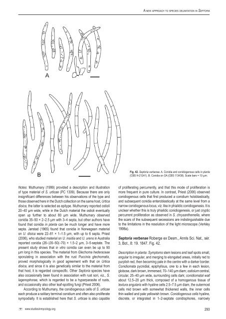
A <strong>new</strong> <strong>approach</strong> <strong>to</strong> <strong>species</strong> <strong>delimitation</strong> <strong>in</strong> Sep<strong>to</strong>riaFig. 42. Sep<strong>to</strong>ria verbenae. A. Conidia and conidiogenous cells <strong>in</strong> planta(<strong>CBS</strong> H-21241). B. Conidia on OA (<strong>CBS</strong> 113438). Scale bars = 10 µm.Notes: Muthumary (1999) provided a description and illustrationof type material of S. urticae (PC 1309). Because there are only<strong>in</strong>significant differences between his observations of the type andthose observed here <strong>in</strong> the Dutch collection on the same host, Urticadioica, the latter is selected as epitype. Muthumary reported ostioli20–40 µm wide, while <strong>in</strong> the Dutch material the ostioli eventuallyopen up further <strong>to</strong> about 80 µm wide. Muthumary observedconidia 35–50 × 2–2.5 µm with 3–4 septa, but other authors havefound that conidia <strong>in</strong> planta can be much longer and have moresepta. Jørstad (1965) found that conidia <strong>in</strong> Norwegean materialon U. dioica were 22–81 × 1–1.5 µm, with up <strong>to</strong> 6 septa. Priest(2006), who studied material on U. <strong>in</strong>sidia and U. urens <strong>in</strong> Australiareported conidia (26–)35–50(–70) × 1.5–2 µm, 3–5-septate. Thepresent study shows that <strong>in</strong> vitro conidia can even be up <strong>to</strong> 90µm long <strong>in</strong> this <strong>species</strong>. The material from Glechoma hederaceaesporulat<strong>in</strong>g <strong>in</strong> association with the rust Pucc<strong>in</strong>ia glechomatis,proved morphologically <strong>in</strong> good agreement with that on Urticadioica, and s<strong>in</strong>ce it is also genetically similar <strong>to</strong> the material fromthat host, it is regarded conspecific. Other Sep<strong>to</strong>ria <strong>species</strong> havealso occasionally been found <strong>in</strong> association with rust sori, viz., S.lagenophorae, which is regarded <strong>to</strong> be a hyperparasite of rusts,and occasionally also other leaf-spott<strong>in</strong>g fungi (Priest 2006).Accord<strong>in</strong>g <strong>to</strong> Muthumary, the conidiogenous cells of S. urticaeeach produce a solitary term<strong>in</strong>al conidium and often also proliferatesympodially. It is established here that S. urticae is also capableof proliferat<strong>in</strong>g percurrently, and that this mode of proliferation ismore frequent <strong>in</strong> pure culture. In contrast, Priest (2006) observedconidiogenous cells that first produced a conidium holoblastically,and subsequent conidia enteroblastically at the same level from anarrow conidiogenous locus, viz. like <strong>in</strong> phialidic conidiogenesis. It isunclear whether this is truly phialidic conidiogenesis, or just crypticpercurrent proliferation as observed <strong>in</strong> S. chrysanthemella, wherethe scars of the subsequent secessions are <strong>in</strong>dist<strong>in</strong>guishable due<strong>to</strong> the limitations <strong>in</strong> the resolution of the light microscope (Verkley1998a).Sep<strong>to</strong>ria verbenae Roberge ex Desm., Annls Sci. Nat., sér.3, Bot., 8: 19. 1847. Fig. 42.Description <strong>in</strong> planta: Symp<strong>to</strong>ms stem lesions and leaf spots small,angular <strong>to</strong> irregular, and merg<strong>in</strong>g <strong>to</strong> elongated areas, <strong>in</strong>itially red <strong>to</strong>purplish red, then becom<strong>in</strong>g pale <strong>in</strong> the centre with a darker border.Conidiomata pycnidial, epiphyllous, one <strong>to</strong> a few <strong>in</strong> each lesion,globose, dark brown, immersed, 70–140 µm diam; ostiolum central,circular, 25–40 µm wide, surround<strong>in</strong>g cells dark; conidiomatal wallabout 12.5–20 µm thick, composed of a homogenous tissue oftextura angularis with hyal<strong>in</strong>e cells 2.5–7.5 µm diam, the outermostcells mid brown with somewhat thickened walls, the <strong>in</strong>ner cellsth<strong>in</strong>-walled and pale yellowish brown. Conidiogenous cells hyal<strong>in</strong>e,discrete, or <strong>in</strong>tegrated <strong>in</strong> 1–2-septate conidiophores, narrowlywww.studies<strong>in</strong>mycology.org293


