Verkley et al.wk (15–18 mm <strong>in</strong> 7 wk), as on MEA, but aerial mycelium denserwith longer hyphae; conidiomatal <strong>in</strong>itials develop<strong>in</strong>g scarcely at thesurface, still sterile after 3 wk, but later on releas<strong>in</strong>g dirty buff <strong>to</strong>pale ochreous droplets of conidial slime. In older colonies on MEAand CHA a grey or greyish white, dense mat of aerial hyphae maycover small or larger sec<strong>to</strong>rs.Conidiomata as <strong>in</strong> vitro, pycnidial, often merged <strong>to</strong> complexstromata, first brownish, then black, glabrous or the surface coveredby short white hyphae; conidiogenous cells as <strong>in</strong> planta, but larger,7.5–20 × 3–5(–6) µm, holoblastic, proliferat<strong>in</strong>g sympodially, nopercurrent proliferation observed; conidia similar <strong>in</strong> shape as <strong>in</strong>planta but mostly 3–7-septate, (45–)55–85(–105) × 4–5(–7) µm.Hosts: Clematis spp.Material exam<strong>in</strong>ed: Austria, Tirol, Ötztal, Brunau, on liv<strong>in</strong>g leaves of Clematisvitalba, 30 July 2000, G. Verkley 1025, epitype designated here <strong>CBS</strong> H-21182“MBT175353”, liv<strong>in</strong>g cultures ex-epitype <strong>CBS</strong> 108983, 108984; same loc.,substr., date, G. Verkley 1026, <strong>CBS</strong> H-21183; same substr., S. Tirol, Eggenthal,Birchabruck, 23 July 1904, J. Kabát, distributed <strong>in</strong> Kabát & Bubák, Fungi imperfectiexsicc. 163, PC 0084599. France, Parc de Lébisey, 27 July 1848, Roberge (?), ‘Col.Desmazieres 1863, no. 8, 448’, isotype PC 0084593; same loc., substr., June 1848,Roberge, PC 0084596; same substr., Paris, Parc de St Cloud, Aug. 1908, Ludwig,PC 0084607; same substr., Fonta<strong>in</strong>ebleau forest, Aug. 1885, PC 0084604; samesubstr., Clères, 27 Aug. 1896 (herb. Mussat), PC 0084598; same substr., Se<strong>in</strong>e-et-Oise, Meudon, 15 Nov. 1844, Roussel (Herb. Roussel), PC 0084594, PC 0084595.Romania, distr. Iaşi, Moldova, Bârnova, same substr., 30 Aug. 1934, T. Săvulescu& C. Sandhu, distributed <strong>in</strong> Săvulescu, Herb. Mycol. Romanicum 24, 1160, PC0084603, 0084608, 0084597.Notes: This is one of the large-spored <strong>species</strong> of Sep<strong>to</strong>ria fromthe genus Clematis. Teterevnikova-Babayan (1987), who studiedcollections from several <strong>species</strong> of Clematis observed, 4–6-septateconidia 60–90 × 3–5 µm. Vanev et al. (1997) reported conidiaas 39–100 × 2.5–4 µm. The type of S. clematidis <strong>in</strong> PC showed4-7-septate conidia 52–78 × 3–3.5 µm, <strong>in</strong> good agreement with theones observed <strong>in</strong> the Austrian material (<strong>CBS</strong> H-21182), which isdesignated above as epitype.The taxonomy of the 15 described <strong>species</strong> of Sep<strong>to</strong>ria onClematis is still unresolved (Sh<strong>in</strong> & Sameva 2004), and wouldcerta<strong>in</strong>ly benefit from study of additional fresh material and cultureswhich could be compared with type material. Sep<strong>to</strong>ria clematidisRoberge is probably dist<strong>in</strong>ct from S. clematidis Pandotra & K.S.M.Sastry, a taxon described on Clematis grata <strong>in</strong> India that should berenamed because it is a later homonym. Accord<strong>in</strong>g <strong>to</strong> Muthumary(1999), the conidia <strong>in</strong> the type of S. clematidis Pan. & Sastry are1–3-septate, 38–66 × 2.5–3 µm, whereas <strong>in</strong> the orig<strong>in</strong>al diagnosisthe conidia are described as “ septate”, 25.6–44.8 (av. 36.3) × 2.3–3.2 (av. 2.7). Two other large-spored <strong>species</strong> are S. jackmanii Ellis& Everh. 1892, which was described from Clematis jackmanii <strong>in</strong>Geneva, New York and, accord<strong>in</strong>g <strong>to</strong> the diagnosis, has conidia 40–70 × 2.5–3 µm (number of septa not given), and also S. williamsiaePriest, based on material on C. aristata <strong>in</strong> Australia, which has(1–)3(–4)-septate conidia 20–45(–55) × (1.5–)2 µm (Priest 2006).Sep<strong>to</strong>ria convolvuli Desm., Annls Sci. Nat., sér. 2, Bot.17:108. 1842. Fig. 14.Description <strong>in</strong> planta: Symp<strong>to</strong>ms leaf lesions circular, s<strong>in</strong>gle orconfluent <strong>to</strong> form irregular extended lesions, pale <strong>to</strong> dark brown,show<strong>in</strong>g one <strong>to</strong> several concentric l<strong>in</strong>es and a dark brown,slightly raised l<strong>in</strong>e or zone delimit<strong>in</strong>g the lesion, visible on bothsides of the leaf. Conidiomata pycnidial, epiphyllous, several <strong>in</strong>each lesion, immersed, subglobose <strong>to</strong> globose, brown <strong>to</strong> black,(65–)90–120(–145) µm diam; ostiolum central, circular <strong>to</strong> irregular,<strong>in</strong>itially 20–40 µm wide, later becom<strong>in</strong>g more irregular and up <strong>to</strong>70 µm wide, surround<strong>in</strong>g cells somewhat darker; conidiomatalwall 10–15 µm thick, composed of a homogenous tissue ofhyal<strong>in</strong>e, angular cells, 2.5–4.5 µm diam, the outermost cells palebrown with slightly thickened walls, the <strong>in</strong>ner cells th<strong>in</strong>-walled.Conidiogenous cells hyal<strong>in</strong>e, discrete, rarely <strong>in</strong>tegrated <strong>in</strong> 1-septateconidiophores, narrowly <strong>to</strong> broadly ampulliform, holoblastic,proliferat<strong>in</strong>g percurrently several times, with <strong>in</strong>dist<strong>in</strong>ct annellationson a relatively elongated neck, or sympodially, 6–10(–17) ×2.5–3.5(–4) µm. Conidia filiform <strong>to</strong> filiform-cyl<strong>in</strong>drical, slightly <strong>to</strong>strongly curved, often elegantly flexuous, attenuated <strong>in</strong> the uppercell <strong>to</strong> a narrowly rounded <strong>to</strong> po<strong>in</strong>ted tip, narrowly truncate at thebase, 1–3(–4)-septate, not constricted around the septa, hyal<strong>in</strong>e,contents m<strong>in</strong>ute oil-droplets and granular material <strong>in</strong> the rehydratedstate, (15–)23–42(–50) × 1.5–2 µm (rehydrated). Sexual morphunknown.Description <strong>in</strong> vitro: Colonies on OA 3–5 mm diam <strong>in</strong> 1 wk (16–20mm <strong>in</strong> 25 d; 40–48 mm <strong>in</strong> 33 d), with an even, glabrous marg<strong>in</strong>,which is colourless, or fa<strong>in</strong>tly salmon due <strong>to</strong> a diffusable pigmentalready visible after 1 wk (but fad<strong>in</strong>g after 3 wk); colonies firstrestricted, conical <strong>to</strong> irregularly pustulate, but later spread<strong>in</strong>g,immersed mycelium <strong>in</strong> the centre becom<strong>in</strong>g first yellowish or citr<strong>in</strong>e,then herbage green or darker olivaceous, surrounded by a morepalid, rosy-buff or pale salmon, later hazel outer zone; pycnidiaalready develop<strong>in</strong>g <strong>in</strong> clusters or radiat<strong>in</strong>g rows at the colonysurface, but they rema<strong>in</strong> scarce, later releas<strong>in</strong>g pale rosy-buff orwhitish droplets of conidial slime; aerial mycelium rema<strong>in</strong><strong>in</strong>g scanty,but <strong>in</strong> the centre it may be well-developed, white, woolly; reverse<strong>in</strong> the centre olivaceous-black <strong>to</strong> olivaceous-grey, surrounded bya first salmon or rosy-buff zone where the diffusable pigment isformed, but this becomes hazel. Colonies on CMA 3–5 mm diam<strong>in</strong> 1 wk [(15–)18–21 mm <strong>in</strong> 25 d; 38–40 mm <strong>in</strong> 33 d], as on OA, butsalmon pigment only fa<strong>in</strong>tly visible after 20 d, the marg<strong>in</strong> becom<strong>in</strong>grosy-buff; centre much darker earlier on, entirely olivaceous-black,numerous black papillate <strong>to</strong> rostrate pycnidia develop<strong>in</strong>g after 21 d,releas<strong>in</strong>g pale whitish <strong>to</strong> buff droplets of conidial slime. Colonies onMEA 2–5 mm diam <strong>in</strong> 1 wk [5–11 mm <strong>in</strong> 25 d; 16–18(– 23) mm <strong>in</strong> 33d], with a ruffled, mostly colourless marg<strong>in</strong> already covered by whiteaerial hyphae after 1 wk; a halo of a diffus<strong>in</strong>g pigment is visible after1 wk, which fades later on; colonies restricted, irregularly pustulateand up <strong>to</strong> 3 mm high after 1 wk, immersed mycelium dark, butmostly <strong>in</strong>visible from above due <strong>to</strong> well-developed, white <strong>to</strong> greyish,dense and short-felted aerial mycelium; black conidiomata alreadydevelop<strong>in</strong>g after 1 wk, releas<strong>in</strong>g large masses of buff conidialslime; reverse mostly sepia <strong>to</strong> isabell<strong>in</strong>e. Some colonies may showa more spread<strong>in</strong>g growth after 2 wk <strong>in</strong> sec<strong>to</strong>rs, that are glabrous,immersed mycelium almost black. Colonies on CHA 3–5 mm diam<strong>in</strong> 1 wk (18–30 mm <strong>in</strong> 25 d; 30–34 <strong>in</strong> 33 d), with an even, glabrous,colourless marg<strong>in</strong>; colonies irregularly pustulate, up <strong>to</strong> 3 mm highafter 1 wk, immersed mycelium colourless <strong>to</strong> pale ochreous, but <strong>in</strong>the centre the surface may be already almost black, while after 25d the entire colony atta<strong>in</strong>s that colour, the larger part covered bywell-developed, low, dense, pure white, later smoke-grey <strong>to</strong> greyolivaceous,felty <strong>to</strong> woolly-floccose, aerial mycelium; conidiomatal<strong>in</strong>itials develop<strong>in</strong>g ma<strong>in</strong>ly <strong>in</strong> the centre after 1 wk; reverse mostlyfawn, but later almost entirely brown-v<strong>in</strong>aceous.Conidiomata s<strong>in</strong>gle, 60–150 µm diam, or merged <strong>to</strong> smallclusters of up <strong>to</strong> 350 µm diam, olivaceous <strong>to</strong> brown, formed mostlyon the agar surface; conidiogenous cells as <strong>in</strong> planta, 6–20 ×2.5–4(–5) µm; conidia as <strong>in</strong> planta, but often some conidia with250
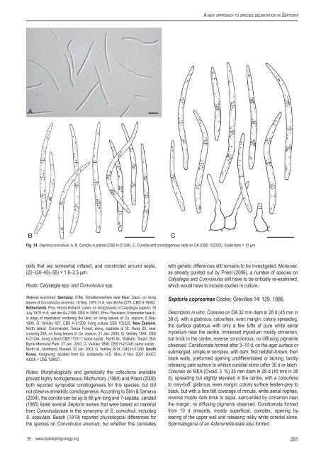
A <strong>new</strong> <strong>approach</strong> <strong>to</strong> <strong>species</strong> <strong>delimitation</strong> <strong>in</strong> Sep<strong>to</strong>riaFig. 14. Sep<strong>to</strong>ria convolvuli. A, B. Conidia <strong>in</strong> planta (<strong>CBS</strong> H-21244). C. Conidia and conidiogenous cells on OA (<strong>CBS</strong> 102325). Scale bars = 10 µm.cells that are somewhat <strong>in</strong>flated, and constricted around septa,(22–)30–45(–55) × 1.8–2.5 µm.Hosts: Calystegia spp. and Convolvulus spp.Material exam<strong>in</strong>ed: Germany, Eiffel, Schalkenmehren near Maar, Daun, on liv<strong>in</strong>gleaves of Convolvulus arvensis, 16 Sep. 1970, H.A. van der Aa 2276, <strong>CBS</strong> H-18082.Netherlands, Prov. Hoord-Holland, Laren, on liv<strong>in</strong>g leaves of Calystegia sepium, 18July 1970, H.A. van der Aa 2198, <strong>CBS</strong> H-18081; Prov. Flevoland, Erkemeder beach,<strong>in</strong> edge of marshland border<strong>in</strong>g the lake, on liv<strong>in</strong>g leaves of Ca. sepium, 8 Sep.1999, G. Verkley 927, <strong>CBS</strong> H-21209, liv<strong>in</strong>g culture <strong>CBS</strong> 102325. New Zealand,North Island, Coromandel, Tairua Forest, along roadside of St. Hway 25, nearcross<strong>in</strong>g 25A, on liv<strong>in</strong>g leaves of Ca. sepium, 21 Jan. 2003, G. Verkley 1844, <strong>CBS</strong>H-21244, liv<strong>in</strong>g culture <strong>CBS</strong> 113111; same substr., North Isl., Waika<strong>to</strong>, Taupiri, BobByrne Memorial Park, 27 Jan. 2003, G. Verkley 1896, <strong>CBS</strong> H-21248; same substr.,North Isl., Northland, Russell, 30 Jan. 2003, G. Verkley 2014, <strong>CBS</strong> H-21245. SouthKorea, Kangnung, isolated from Ca. soldanella, H.D. Sh<strong>in</strong>, 8 Nov. 2007, KACC43226 = <strong>CBS</strong> 128627.Notes: Morphologically and genetically the collections availableproved highly homogeneous. Muthumary (1999) and Priest (2006)both reported sympodial conidiogenesis for this <strong>species</strong>, but didnot observe annellidic conidiogenesis. Accord<strong>in</strong>g <strong>to</strong> Sh<strong>in</strong> & Sameva(2004), the conidia can be up <strong>to</strong> 68 µm long and 7-septate. Jørstad(1965) listed several Sep<strong>to</strong>ria names that were based on materialfrom Convolvulaceae <strong>in</strong> the synonymy of S. convolvuli, <strong>in</strong>clud<strong>in</strong>gS. septulata. Beach (1919) reported physiological differences forthe <strong>species</strong> on Convolvulus arvensis, but whether this correlateswith genetic differences still rema<strong>in</strong>s <strong>to</strong> be <strong>in</strong>vestigated. Moreover,as already po<strong>in</strong>ted out by Priest (2006), a number of <strong>species</strong> onCalystegia and Convolvulus still have <strong>to</strong> be critically re-exam<strong>in</strong>ed,which would have <strong>to</strong> <strong>in</strong>clude studies <strong>in</strong> culture.Sep<strong>to</strong>ria coprosmae Cooke, Grevillea 14: 129. 1886.Description <strong>in</strong> vitro: Colonies on OA 32 mm diam <strong>in</strong> 28 d (45 mm <strong>in</strong>38 d), with a glabrous, colourless, even marg<strong>in</strong>; colony spread<strong>in</strong>g,the surface glabrous with only a few tufts of pure white aerialmycelium near the centre, immersed mycelium mostly c<strong>in</strong>namon,but brick <strong>in</strong> the centre, reverse concolorous; no diffus<strong>in</strong>g pigmentsobserved. Conidiomata formed after 3–10 d, on the agar surface orsubmerged, simple or complex, with dark, first reddish-brown, thenblack walls, preformed open<strong>in</strong>g undifferentiated or lack<strong>in</strong>g, tardilyreleas<strong>in</strong>g pale salmon <strong>to</strong> whitish conidial slime (after 30 d or later).Colonies on MEA (Oxoid, 3 %) 35 mm diam <strong>in</strong> 28 d (45 mm <strong>in</strong> 38d), spread<strong>in</strong>g but slightly elevated <strong>in</strong> the centre, with a colourless<strong>to</strong> rosy-buff, glabrous, even marg<strong>in</strong>; colony surface leaden-grey <strong>to</strong>black, but with a f<strong>in</strong>e felt coverage of m<strong>in</strong>ute, white aerial hyphae,reverse mostly dark brick <strong>to</strong> sepia, surrounded by c<strong>in</strong>namon nearthe marg<strong>in</strong>; no diffus<strong>in</strong>g pigments observed. Conidiomata formedfrom 10 d onwards, mostly superficial, complex, open<strong>in</strong>g bytear<strong>in</strong>g of the upper wall and releas<strong>in</strong>g milky white conidial slime.Sperma<strong>to</strong>gonia of an Asteromella-state also formed.www.studies<strong>in</strong>mycology.org251