

A new approach to species delimitation in Septoria - CBS - KNAW
A new approach to species delimitation in Septoria - CBS - KNAW
A new approach to species delimitation in Septoria - CBS - KNAW

Create successful ePaper yourself
Turn your PDF publications into a flip-book with our unique Google optimized e-Paper software.
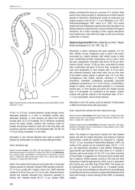
Verkley et al.already considered this name as a synonym of S. heraclei. Otherauthors have mostly accepted S. heracleicola as a further Sep<strong>to</strong>ria<strong>species</strong> on Heracleum, describ<strong>in</strong>g the conidia as cont<strong>in</strong>uous andrang<strong>in</strong>g roughly <strong>in</strong> size 20–40 × 1–2 µm (Radulescu et al. 1973,Teterevnikova-Babayan 1987, Vanev et al. 1997). Four furtherSep<strong>to</strong>ria and two Rhabdospora <strong>species</strong> have been described <strong>in</strong> theliterature based on material found on various members of the genusHeracleum, all of which accord<strong>in</strong>g <strong>to</strong> their orig<strong>in</strong>al descriptionshave conidia more or less with<strong>in</strong> this range, so with much narrowerconidia than S. heraclei.Sep<strong>to</strong>ria hypochoeridis Petrov, Materialy po mikol. i fi<strong>to</strong>pat.Rossii (Len<strong>in</strong>grad) 6 (1): 55. 1927. Fig. 22.Description <strong>in</strong> planta: Symp<strong>to</strong>ms leaf spots scattered, 2–5 mmdiam, def<strong>in</strong>ite, circular, hologenous, grey <strong>to</strong> white <strong>in</strong> the centre,surrounded by a slightly elevated, dark reddish purple or blackzone. Conidiomata pycnidial, hypophyllous, one <strong>to</strong> a few <strong>in</strong> eachleaf spot, (sub)globose, immersed, dark brown, 60–95 µm diam;ostiolum central, circular, 15–28 µm diam, surrounded by darkercells; conidiomatal wall about 10–20 µm thick, composed of anouter layer isodiametric or more irregular cells, 5–10 µm diam,with somewhat thickened, pale brown walls, and an <strong>in</strong>ner layerof th<strong>in</strong>-walled, hyal<strong>in</strong>e angular <strong>to</strong> globose cells, 4.5–7 µm diam.Conidiogenous cells hyal<strong>in</strong>e, discrete, cyl<strong>in</strong>drical, or broadlyampulliform, holoblastic, proliferat<strong>in</strong>g sympodially, percurrentproliferation not observed, 8–15 × 3.5–4(–5.5) µm. Conidia filiform,straight <strong>to</strong> slightly curved, attenuated gradually <strong>to</strong> a somewhatpo<strong>in</strong>ted apex, or more abruptly just above the broadly truncatebase, 0–1(–2)-septate, not constricted at the septum, hyal<strong>in</strong>e,contents with granular material <strong>in</strong> the rehydrated state, 15–24 ×1–1.5 µm (rehydrated). Sexual morph unknown.Fig. 21. Sep<strong>to</strong>ria heraclei, conidia and conidiogenous cells <strong>in</strong> planta (<strong>CBS</strong> H-21224).Scale bars = 10 µm.10–25 × 5–7(–8) µm. Conidia cyl<strong>in</strong>drical, usually strongly curved,attenuated gradually <strong>to</strong> a blunt <strong>to</strong> somewhat po<strong>in</strong>ted apex,attenuated gradually, or more abruptly just above the broadlytruncate base, (0–)1–2(–4)-septate, not or <strong>in</strong>dist<strong>in</strong>ctly constrictedaround the septa, hyal<strong>in</strong>e, contents with numerous small oildropletsand granular material <strong>in</strong> each cell <strong>in</strong> the liv<strong>in</strong>g state, withamorphous granular contents <strong>in</strong> the rehydrated state, 40–55(–70)× 4–6 µm (liv<strong>in</strong>g; rehydrated, 3–5 µm wide).Description <strong>in</strong> vitro: Several attempts were made <strong>to</strong> isolate this<strong>species</strong> but unfortunately no conidia survived after germ<strong>in</strong>ation.Hosts: Heracleum spp.Material exam<strong>in</strong>ed: Austria, Tirol, Ötztal, Ötz near Habichen, on liv<strong>in</strong>g leaves ofHeracleum sphondylium, 24 July 2000, G. Verkley 1002, <strong>CBS</strong> H-21186. Netherlands,Prov. Limburg, Gulpen, near S<strong>to</strong>khem, on liv<strong>in</strong>g leaves of H. sphondylium, 28 June2000, G. Verkley 957, <strong>CBS</strong> H-21224; same substr., Prov. Limburg, upper edge ofSavelsbos, G. Verkley 959, <strong>CBS</strong> H-21225.Notes: The conidia of this fungus are much wider than <strong>in</strong> most otherSep<strong>to</strong>ria <strong>species</strong> on Apiaceae. Jørstad (1965) reported conidia 35–57 × 3–5 µm, usually with 1 septum. Vanev et al. (1997) observedconidia up <strong>to</strong> 85 µm long, and 1.8–3.5 µm wide. Sep<strong>to</strong>ria heracleipalmatiwas orig<strong>in</strong>ally described from Heracleum palmatum <strong>in</strong>Greece, with 1-septate conidia, 50–70 × 3 µm. Jørstad (1965)Description <strong>in</strong> vitro: No cultures could be obta<strong>in</strong>ed. Conidia placedon MEA and OA died shortly after germ<strong>in</strong>ation.Hosts: Hypochoeris radicata and other Hypochoeris spp.Material exam<strong>in</strong>ed: New Zealand, North Island, Taupo distr., Tongariro Nat. Park,Taurewa, along road 47, on decay<strong>in</strong>g leaf base of Hypochoeris radicata, 25 Jan.2003, G. Verkley 1871, <strong>CBS</strong> H-21234.Additional material exam<strong>in</strong>ed: New Zealand, North Island, Taupo distr., Lake Taupo,shorel<strong>in</strong>e E of Motutaiko Island, on liv<strong>in</strong>g leaves of Crepis capillaris, 25 Jan. 2003,G. Verkley 1870, <strong>CBS</strong> H-21235.Notes: The material on Hypochoeris radicata from New Zealandagrees well with the orig<strong>in</strong>al description and draw<strong>in</strong>g of Sep<strong>to</strong>riahypochoeridis; conidia are reported as cont<strong>in</strong>uous <strong>to</strong> 1-septate,19–22 × 1.5 µm. Accord<strong>in</strong>g <strong>to</strong> Teterevnikova-Babayan (1987), theconidia of this <strong>species</strong> can be somewhat larger, 20–25 × 1.5–2µm, and Hypochoeris grandiflora is also <strong>in</strong>fected. Rhabdosporahypochoeridis was described from dead stems of H. radicata <strong>in</strong>Germany, with curved conidia, 16–30 × 0.6–1 µm, which, accord<strong>in</strong>g<strong>to</strong> Priest (2006), is suggestive of a Phomopsis with β-conidia ratherthan a Sep<strong>to</strong>ria. Another <strong>species</strong> ocurr<strong>in</strong>g on this host and otherAsteraceae is Sep<strong>to</strong>ria lagenophorae, which occurs <strong>in</strong> associationwith Pucc<strong>in</strong>ia spp. and other fungi (Priest 2006). This fungus canbe dist<strong>in</strong>guished from S. hypochoeridis by 1–2-septate conidia,15–32 µm long, and conidiogenous cells which are not proliferat<strong>in</strong>gsympodially but produce successive conidia enteroblastically at thesame level through a narrow open<strong>in</strong>g (Priest 2006), so appear<strong>in</strong>gphialidic.262


